| Parasite | Anisakis sp. |
|---|---|
| Taxonomy | Nematoda, Secernentea, Anisakidae |
| Host | European hake (Merluccius merluccius) |
| Infection site | Muscle |
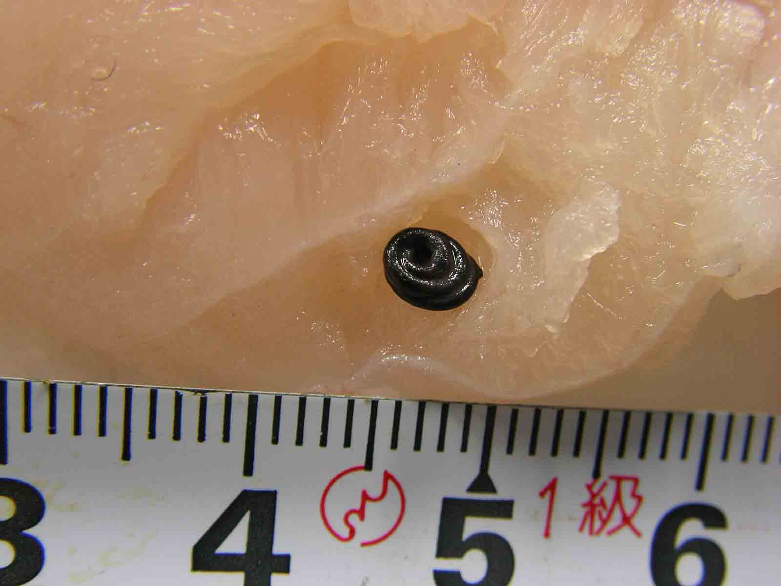
| Clinical sign | A coiled black filiform worm is observed in a fillet of European hake imported from Chile (Fig. 1). |
| Parasitology | A black membrane surrounding the parasite is formed by the host reaction. A larva of Anisakis sp. appears inside the membrane (Fig. 2). This parasite is probably an Anisakid nematode since it lacks caecum (Fig. 4), possesses boring tooth (Fig 3) and mucron (tail spine) at the anterior and posterior end, respectively. This parasite is different from the typical A. simplex s. s. because of the difference in RFLP pattern of the parasite from A. simplex in Japan. |
| Pathology | Pathogenicity to fish is low. |
| Health hazard | This parasite probably cause the human anisakiasis. |
| Diagnosis | This parasite can be morphologically diagnosed as L3 larva of Anisakis type I based on the presence of mucron at the blunt posterior end, oesophagous and simple, long intestine but the absence of caecum (Koyama, 1974). However, Anisakid larvae cannot be classified into more detail by morphology. Therefore, sequences of rRNA or RFLP mapping are used to identify them (Abe et al., 2006; Valentini et al., 2006; Umehara, et al., 2006). |
| References | Abe, N., K. Tominaga
and I. Kimata (2006): Usefulness of PCR-restriction fragment length polymorphism
analysis of the internal transcribed spacer region of rDNA for identification
of Anisakis simplex complex. Japan. J. Infect. Dis., 59, 60-62. Koyama, T (1974): I. Anisakidae larvae. 1. Morphology and classification. Fish and anisakis (The Japanese Society of Fisheries Science), Koseisha koseikaku, pp.9-19. (In Japanese) Umehara, A., Y. Kawakami, T. Matsui, J. Araki and A. Uchida (2006): Molecular identification of Anisakis simplex sensu stricto and Anisakis pegreffii (Nematoda : Anisakidae) from fish and cetacean in Japanese waters. Prasitol. Int., 55, 267-271. Valenti, A., S. Mattiucci, P. Bondanelli, S. C. Webb, A. A. Mignucci-Giannone, M. M. Colom-Llavina and G. Nascetti (2006): Genetic relationships among Anisakis species (Nematoda: Anisakidae) inferred from mitochondrial COX2 sequences, and comparison with allozyme data. J. Parasitol., 92, 156-166. |
Fig. 3. The anterior end of the Anisakis.

Fig. 4. The ventriculus of Anisakis sp. Note the absence of caecum.
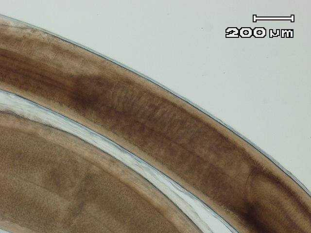
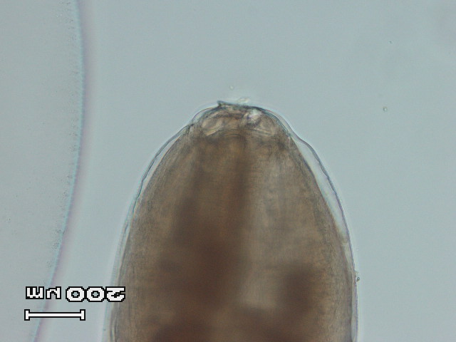
Fig. 2. A black membrane surrounding the worm.
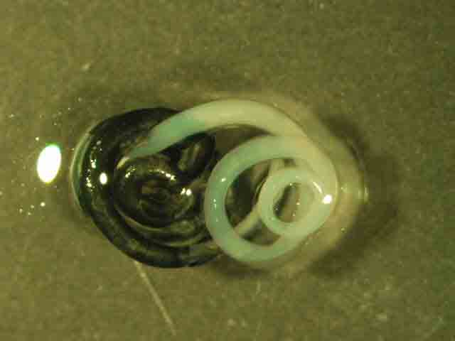
Fig. 1. Anisakis found in the muscle of hake.