| Parasite | Anisakis pegreffii |
|---|---|
| Taxonomy | Nematoda, Secernentea, Anisakidae |
| Hosts | Marine fishes including greater amberjack (Seriola dumerili), chicken grunt (Parapristipoma trilineatum), chub mackerel (Scomber japonicus), blue mackerel (Scomber australasicus), etc. |
| Infection site | Body cavity, visceral organs, muscle |
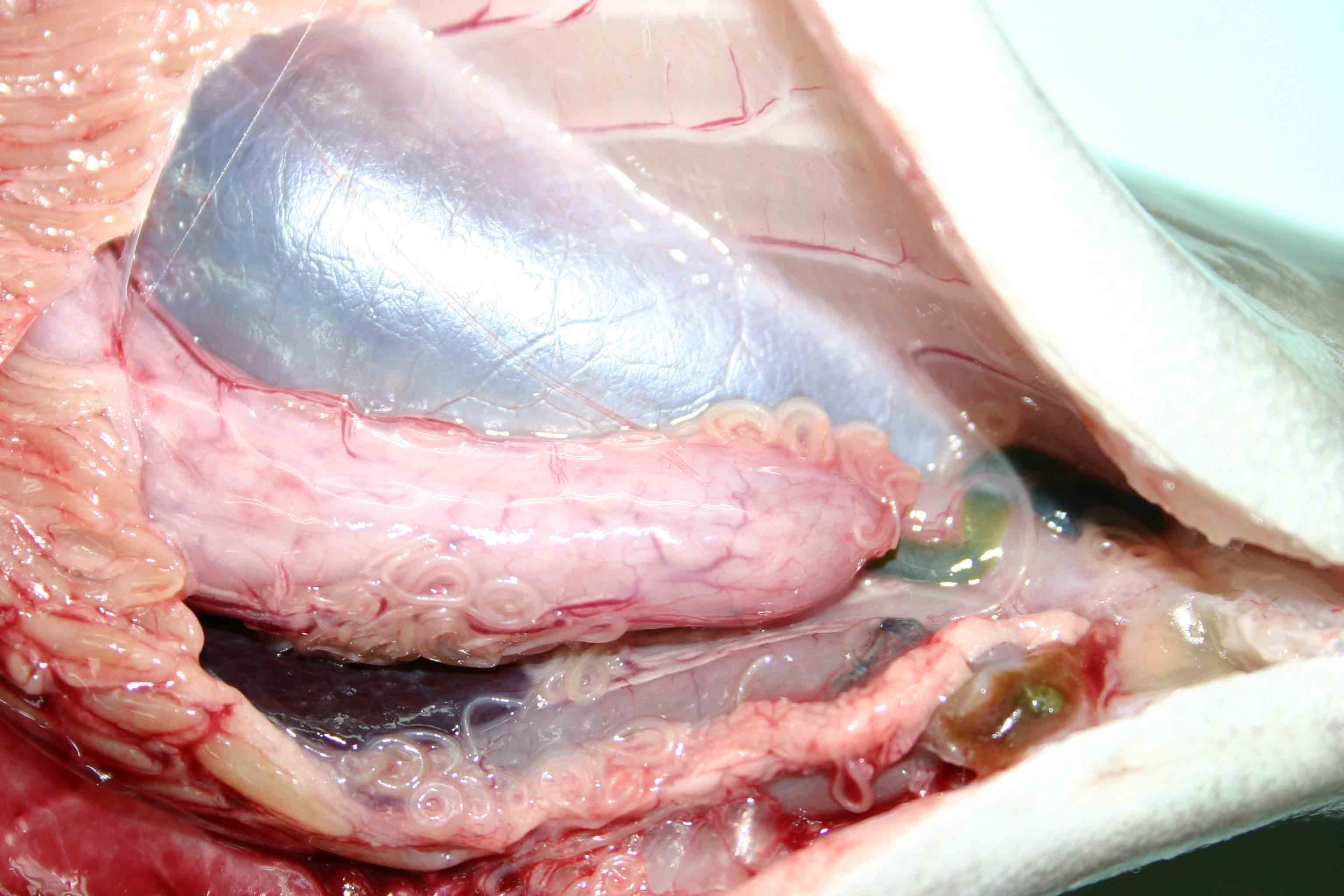
| Clinical sign | Infected fish exhibits no external abnormality. Coiled filiform parasites (ca. 1-3 cm) are observed in the body cavity and on the liver or stomach. In some fishes, this parasite infects the muscle. |
| Parasitology | Final host is mainly whales, and the parasite develops to the adult inside their stomachs followed by the egg production. Released larva hatched from the egg is ingested by crustacean, e. g. euphausiid. Fishes are infected by eating such krill. Previously, it was considered that Anisakis type I, of which L3 larva causes anisakiasis, is only Anisakis simplex. However, recent studies revealed that type I is composed of A. simplex, A. simplex C, A. pegreffii, A. typical and A. ziphidarum (Mattiucci et al., 2002). In a limited sense (sensu stricto), ‘A. simplex’ means only the one species A. simplex, meanwhile, in the broad sense (sensu lato), ‘A. simplex’ includes A. simplex C and A. pegreffii. |
| Pathology | Pathogenicity to fish may be low. However, some pathological effects are reported in greater amberjack, e. g. a deformed and shortened stomach (Yoshinaga et al., 2006). |
| Health hazard | A live parasite, a causative agent of anisakiasis, penetrates into the gut wall of human and causes the acute gastroenteritis, viz. violent abdominal pain, nausea and vomiting. Therefore, be careful when the host fishes is consumed raw. Heating or freezing to -20 deg C for 24 hours is an effective method of killing the parasite. |
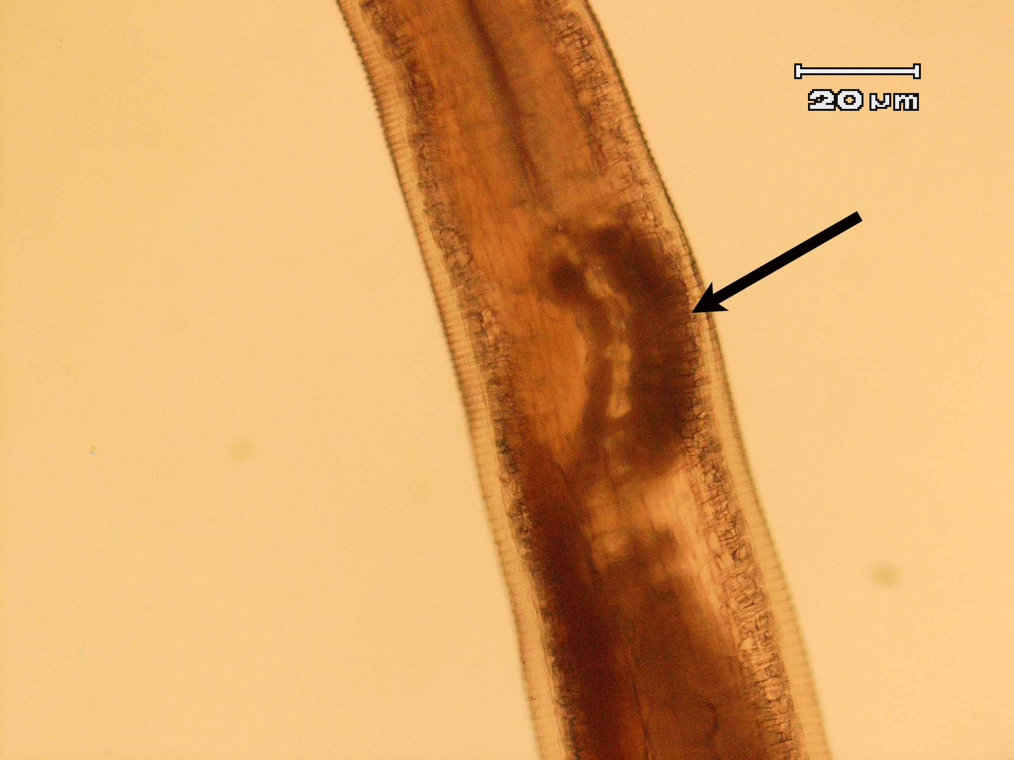
| Diagnosis | L3 larva of Anisakis type I has boring tooth at the anterior end, oesophagous, ventriculus and intestine but no caecum. The larva of type I is distinguished from that of type II by the presence of the mucrons (tail spine) and the blunt posterior end of type I (Koyama, 1974). A. pegreffii is distinguished from A. simplex sensu stricto in the length of ventriculus (the former one is shorter). However, it is difficult to classify among type I only based on morphology. Therefore, sequences of rRNA or PCR-restriction fragment length polymorphism (RFLP) mapping are used to identify them (Abe et al., 2006; Valentini et al., 2006; Umehara, et al., 2006). |
| Other information | In 2005, juvenile greater amberjack imported from China to Japan were found to be infested with Anisakis type I larvae (probably A. pegreffii). It is considered that the amberjack were infected with the parasite in China before importation, where they were fed raw fish harboring the parasite (Yoshinaga et al., 2006). |
| References | Abe, N., K. Tominaga
and I. Kimata (2006): Usefulness of PCR-Restriction Fragment Length
Polymorphism Analysis of the Internal Transcribed Spacer Region of rDNA for
Identification of Anisakis simplex
Complex. Japan. J. Infect. Dis., 59, 60-62.
Koyama, T (1974): I. Anisakidae larvae. 1. Morphology and classification. Fish and anisakis (The Japanese Society of Fisheries Science), Koseisha koseikaku, pp.9-19. (In Japanese) Mattiucci, S., L. Paggi, G. Nascetti, C. P. Santos, G. Costa, A. P. Di Beneditto, R. Ramos, M. Argyrou, R. Cianchi and L. Bullini (2002): Genetic markers in the study of Anisakis typica (Diesing, 1860): larval identification and genetic relationships with other species of Anisakis Dujardin, 1845 (Nematoda: Anisakidae). System. Parasitol., 51, 159-170. Umehara, A., Y. Kawakami, T. Matsui, J. Araki and A. Uchida (2006): Molecular identification of Anisakis simplex sensu stricto and Anisakis pegreffii (Nematoda : Anisakidae) from fish and cetacean in Japanese waters. Prasitol. Int., 55, 267-271. Valentini, A., S. Mattiucci, P. Bondanelli, S. C. Webb, A. A. Mignucci-Giannone, M. M. Colom-Llavina and G. Nascetti (2006): Genetic relationships among Anisakis species (Nematoda: Anisakidae) inferred from mitochondrial COX2 sequences, and comparison with allozyme data. J. Parasitol., 92, 156-166. Yoshinaga, T., R. Kinami, K. A. Hall and K. Ogawa (2006): A preliminary study on the infection of Anisakid larvae in juvenile greater amberjack Seriolae dumerili imported from China to Japan as mariculture seedlings. Fish Pathol., 41, 123-126. |
Fig. 2. The ventriculus (arrow) ofAnisakis pegreffi, which is shorter
than that of A. simplex.
Fig. 1. Anisakis pegreffi found in the body cavity of greater amberjack
(Photos by T. Yoshinaga (1) and Kark Marx Quiazon (2))