
| Parasite | Digramma interrupta |
|---|---|
| Taxonomy | Plathyhelminthes, Cestoda, Pseudophyllidea, Ligulidae |
| Hosts | Crucian carp (Carassius auratus), rosyface dace (Tribolodon ezoe) |
| Infection site | Peritoneal cavity |
| Clinical signs | Diseased fish exhibit abdominal swelling and lethargy (Figs. 1 and 2). A white vermiform parasite (tapeworm) occupies the peritoneal cavity (Fig. 3). |
| Parasitology | The body is 1.5 cm wide and over 1 m long (Fig. 4). Fish (the second intermediate host) is infected with larvae (plerocercoids). A scolex and proglottids are not observed in the larvae (Awakura, 2006). The first intermediate host and the definitive host for this parasite are copepod and fish-eating birds, respectively. The parasite matures and produces eggs in the digestive tract of birds. |
| Pathology | The infection inhibits the development of host’s gonad, leading to castration of the host (Awakura et al., 1976). |
| Health hazard | Since this parasite is not infectious to human, it is harmless in food hygiene. Anecdotally, this parasite is called as “marine macaroni (maccheroni di mare)” and provided for human consumption in some areas of Italy. |
| Diagnosis | The parasite can be visually observed in the peritoneal cavity. |
| Other information | An adult of the parasite can be experimentally obtained by incubating the larva in horse serum for 2-3 days. There are no effective methods for control of this disease. |
| References | Awakura, T. (2006): Digramma infection. New atlas of fish diseases (eds. by Hatai, K. and K. Ogawa), Midori
Shobo, p. 123. (In Japanese) Awakura, T., H. Tonosaki and T. Ito (1976): Ligulosis of cyprinid in the lakes of Hokkaido, Japan. Sci. Rep. Hokkaido Fish Hatchery, 31, 67-81. (In Japanese) |
(Photos by T. Awakura)

Fig. 3. D. interrapta from rosyface dace.
Fig. 2. Digramma in the peritoneal cavity of rosyface dace.
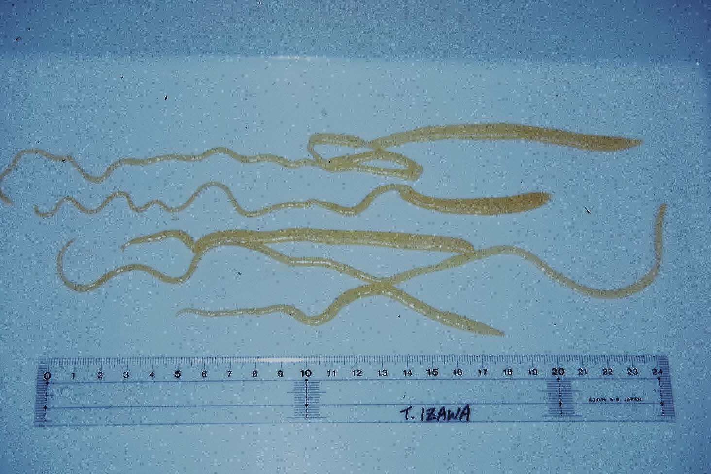
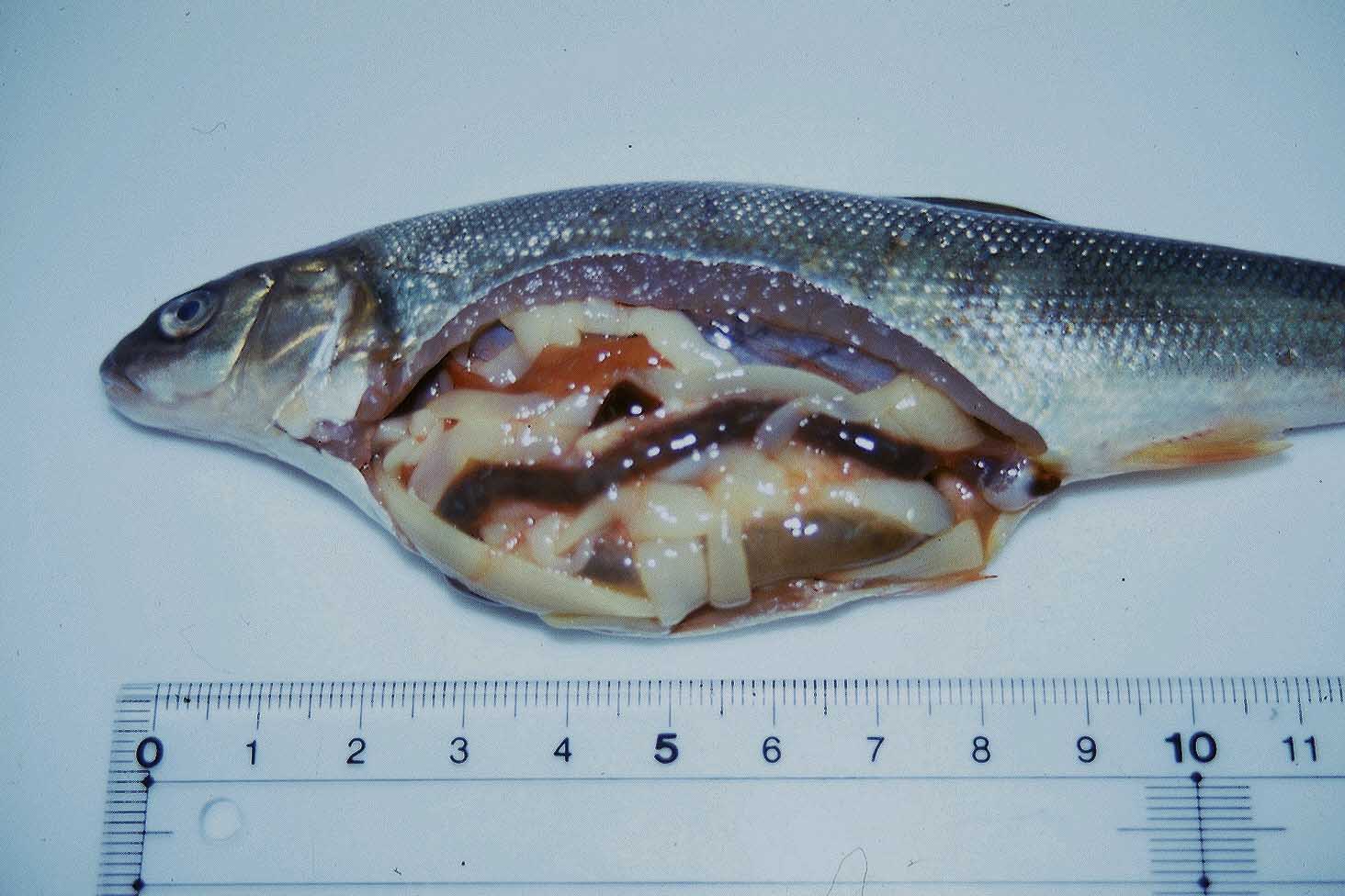
Fig. 1. Crucian carp showing the abdominal distension.