| Parasite | Glugea epinephelusis |
|---|---|
| Taxonomy | Microspora, Microsporea |
| Host | Hong Kong grouper (Epinephelus akaara) |
| Infection site | Abdominal cavity |
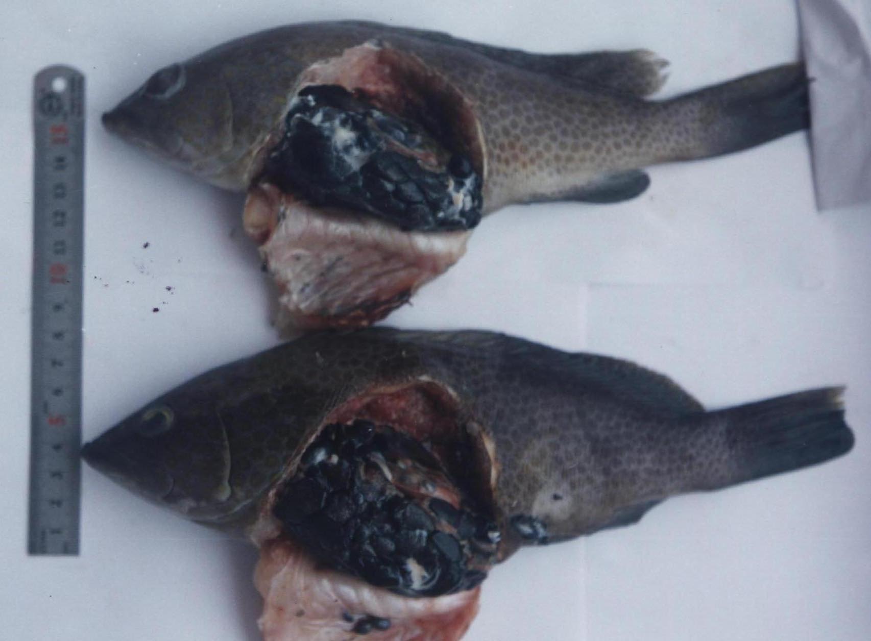
| Clinical sign | Many black amorphous cysts (several mm to 1 cm) are observed in the abdominal cavity (Fig. 1 and 2). |
| Parasitology | The cyst is a host-parasite complex, called asxenoma. The parasite develops and finally produces numerous spores inside. A spore is ellipsoidal and 4-5 mm in length (Fig. 3). |
| Pathology | The reason of black appearance of the xenoma is unknown, but may be caused by melanin deposition by the host. Mortality due to heavy infection may occur. |
| Health hazard | Since this parasite is not infectious to human, it is harmless in food hygiene. |
| Diagnosis | When black cysts are observed inside the peritoneal cavity, squash the cysts and check the spores by wet-mount. If almost all spores are disrupted, smear the sample and stain by Uvitex 2B followed by a fluorescent microscopic observation. The stained spores emit blue fluorescence under UV radiation. |
| Other information | Outbreaks of this disease occurred in fish farms of Hong Kong grouper in GuangDong Province in China (Wu et al., 2005; Zhang et al., 2004). Though infected fish produces a specific antibody to the parasite spores, the intensity of infection (number of cysts) is not related to the antibody level (Zhang et al., 2005). |
| References | Wu, H. B., Y. S. Wu
and Z. H. Wu (2005): Occurrence of a new microsporidium in the abdominal cavity
of Epinephelus akaara. Acta Hydrobiol. Sin. 29, 150-154. Zhang, J. Y., Y. S. Wu, Y. S. Lu and J. G.Wang (2004): Advance in research of fish microsporidia. Acta Hydrobiol. Sin. 28, 563-568. Zhang, J. Y., Y. S. Wu, H. B. Wu, J. G. Wang, A. H. Li and M. Li (2005): Humoral immune responses of the grouper Epinephelus akaara against the microsporidium Glugea epinephelusis. Dis. Aquat. Org., 64, 121-126. |

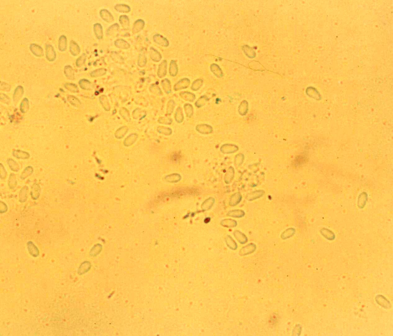
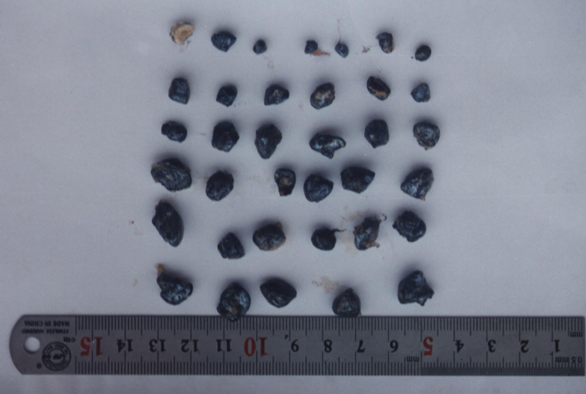
Fig. 2. Xenomas of Glugea from the grouper.
Fig. 1. Hong Kong grouper infected with G. epinephelusis.
Fig. 3. Fresh sporers of G. epinephelusis.
(Photos by Jin-Yong Zhang)