| Parasite | Glugea plecoglossi |
|---|---|
| Taxonomy | Microspora, Microsporea |
| Hosts | Ayu (Plecoglossus altivelis), rainbow trout (Oncorhynchus mykiss) |
| Infection site | Peritoneal cavity |
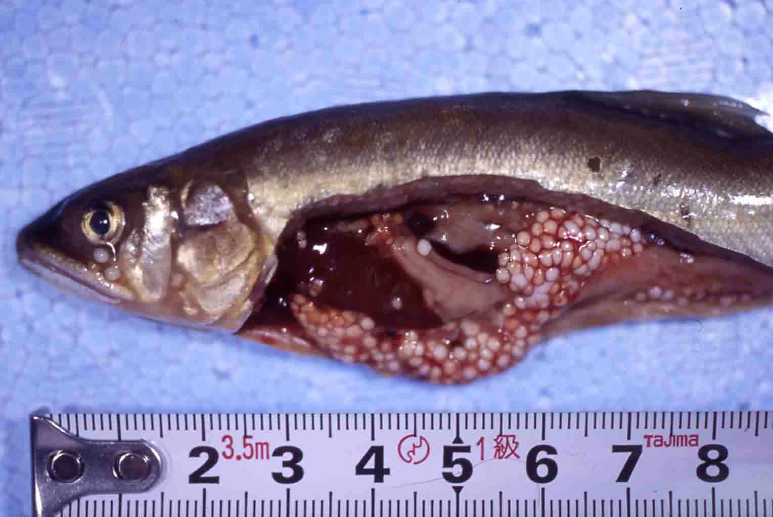
| Clinical signs | Infected fish exhibits no significant external abnormalities, while the abdomen is filled with numerous Glugea cysts (ca. 4-5 mm) (Fig. 1). On the other hand, in heavily infected fry of ayu, cysts are easily observed by transmitted light even without dissection (Fig. 2). |
| Parasitology | Many spores are produced inside xenomas (a complex formed by a host cell and a parasite) (Fig. 3). A spore (average length 5.8 mm; average width 2.1 mm) is ellipsoidal. |
| Pathology | Spores are ingested by the host's phagocytic cells and develop inside the cells. These cells begin to hypertrophy, take a nutrition from the host and result in xenomas. When the xenoma establishes on the wall of the peritoneal cavity, it is encased by the host's connective tissues. The parasite proliferates, and fills up the xenoma. Xenoma, a few mm (up to 5 mm) in size, is visually observed as a cyst (Fig. 4). In a heavy infection, diseased fish sometimes exhibits ascites and emaciates, but few fish are led to the death. |
| Health hazard | Since this parasite is not infectious to human, it is harmless in food hygiene. |
| Diagnosis | Observe the cysts in the peritoneal cavity. For confirmation, check the spores by the wet-mount of xenomas. The sample should be smeared and stained by Uvitex 2B followed by a fluorescent microscopic observation. The stained spores emit blue fluorescence under UV radiation. |
| Other information | Since the glugeosis occurrs when fry of ayu from the Lake Biwa is maintained with well water, Glugea plecoglossi is endemic in the Lake Biwa. However, infection cycle of this disease is uncertain because no infected wild ayu was found in Lake Biwa (Takahashi and Ogawa, 1997). Although this disease has been also reported in other areas (Takahashi, 1981), it is unknown that infection cycle is maintained such areas. Because glugeosis in fry during the seed production may be derived from its parents, check the gonads not to introduce the pathogen. To prevent the disease effectively, maintain the fish at 28-29 C. for 5 days, then return to the normal temp. for 7 days, and raise to 28-29 C. for 5 days again before the fish develops the disease, e. g. just after the introduction of wild seedlings. Take care of other diseases or progress of the sexual maturation when maintaining at high temperature. |
| References | Takahashi, S. (1981): Studies
on Glugea infection of ayu, Plecoglossus altivelis. Bull. Shiga Pref. Fish. Exp. Station, 34, 1-81.(In Japanese) Takahashi, S. and K. Ogawa (1997): Efficacy of elevated water temperature treatment of ayu infected with the microsporidian Glugea plecoglossi. Fish Pathol., 32, 193-198. |
Fig. 2. Artificially produced seedlings of ayu infected
with Glugea
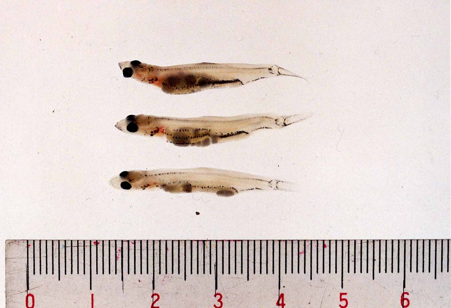
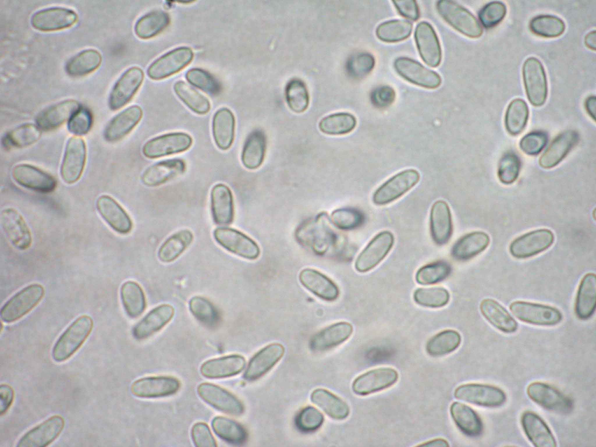
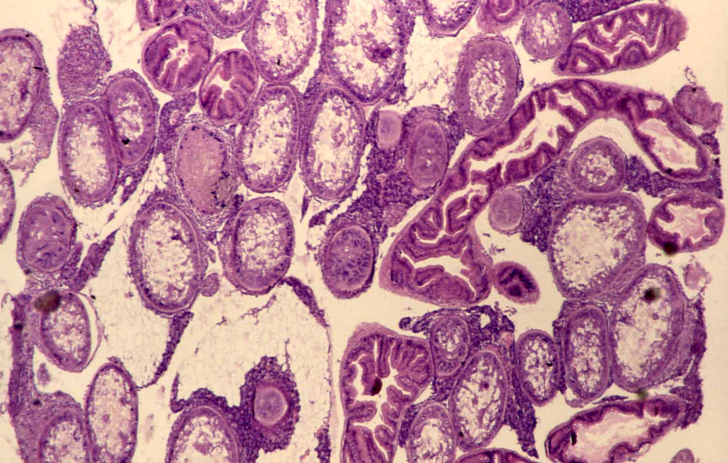
Fig. 3. Fresh spores of G. plecoglossi
Fig. 4. Histology of xenomas in the peritoneal cavity.

Fig. 1. Many xenomas are observed in the body cavity of ayu.