
| Parasite | Hoferellus carassii |
|---|---|
| Taxonomy | Myxozoa, Myxosporea, Bivalvulida |
| Hosts | Crucian carp, goldfish (Carassius auratus) |
| Disease name | Kidney enlargement disease |
| Infection site | Kidney |
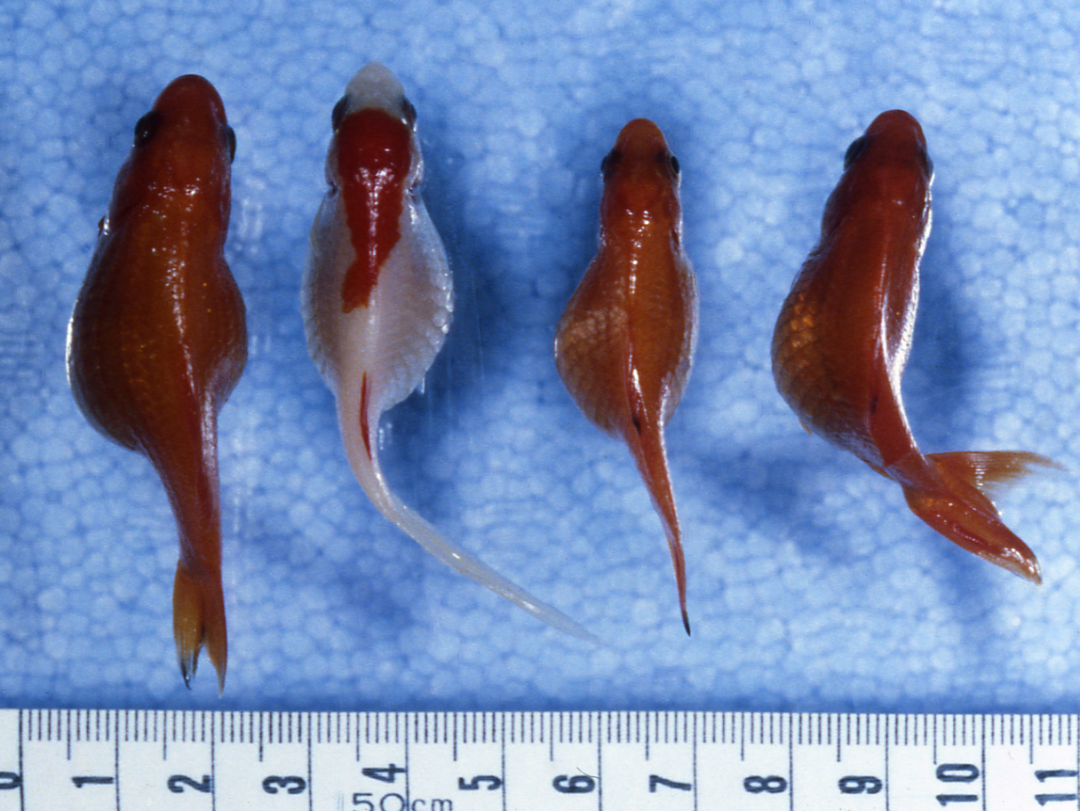
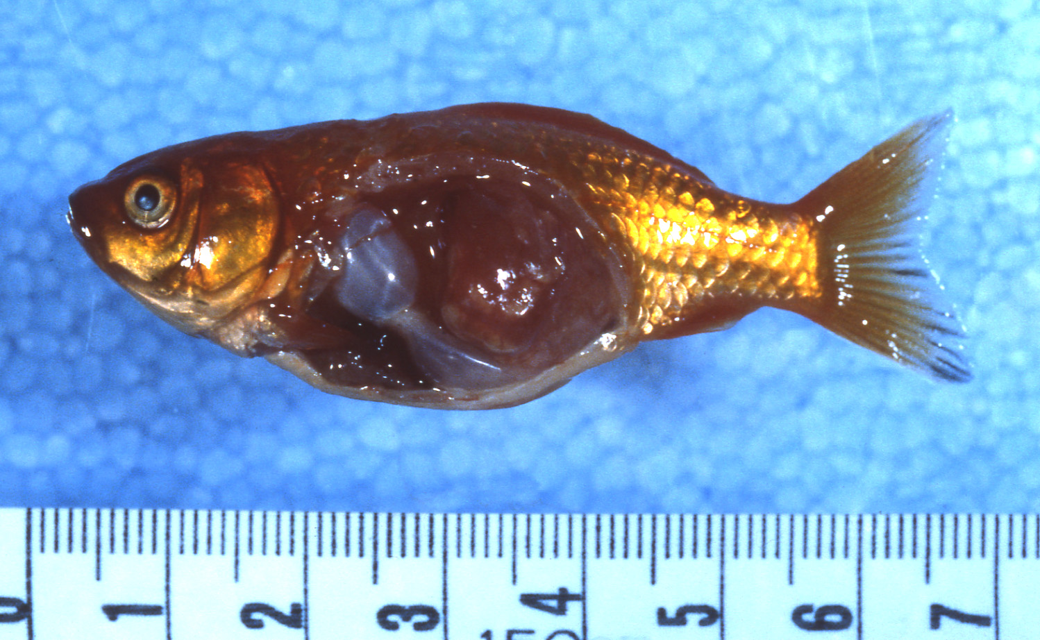
| Clincal signs | This disease is characterized by the abdominal distension. The body of diseased fish often curves laterally because the abdomen swells toward one side (Fig. 1). The enlarged kidney presses the visceral organs, for example swim bladder (Fig. 2). |
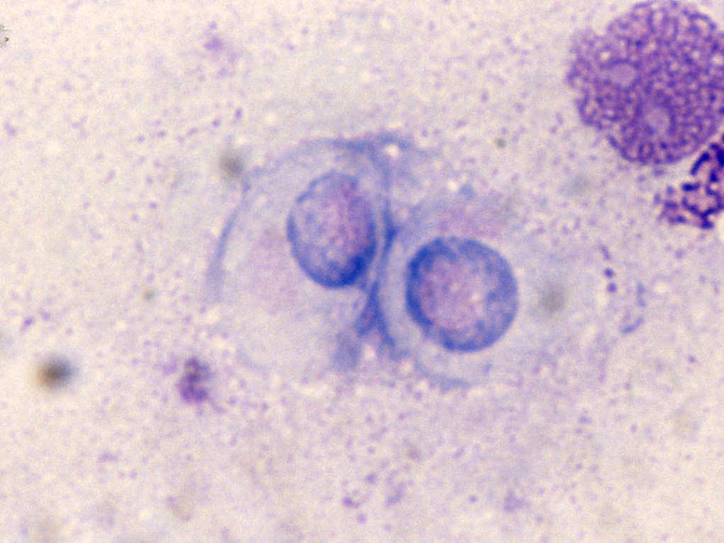
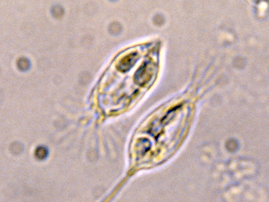
| Parasitology | In October to November, trophozoites (ca. 10 mm in diameter) proliferate in the epithelial cell of the renal tubule (Fig. 3). The parasite develops to a multicellular plasmodium (several tens of mm), in which tertiary and quartenery cells are differentiated. Plasmodium is released to the lumen, and spores are formed in the next spring, when the water temperature increases (Yokoyama et al., 1990a). Spores are released outside the host, and ingested by the oligochaetes followed to actinosporean development (Yokoyama et al., 1993, Trouillier et al., 1996). A miter-like spore (length ca. 12 mm; width ca. 6 mm) has 2 polar capsules (length ca. 4 mm) and bristles at the posterior end. |
| Pathology | Division of the proliferative stages of the parasite is followed by multiplication of the infected host epithelial cells, resulting in hypertrophy of the kidney (Yokoyama et al., 1990a). Even after the release of spores outside the host, the kidney remains enlarged. However, the kidney functions are not so impaired because only one kidney is infected. Diseased fish would become exhausted and get secondary infection of bacteria resulting from the swimming disability. |
| Health hazard | Since this parasite is not infectious to human, it is harmless in food hygiene. |
| Diagnosis | Mature spores can be observed in a wet-mount preparation of the kidney in spring or later. However, during the peak season of disease outbreak (autumn to winter), spores are not produced. At that time, the characteristic morphology of trophozoites (multicellular and multinuclear plasmodium) should be checked in a Giemsa or Diff-Quik stained smear. |
| Other information | This disease was first noticed in 1963 at goldfish farms in Tokyo, and then spread to other areas. A high temperature treatment at 30 C (Yokoyama, 2002) or an oral administration of fumagillin (Yokoyama et al., 1990b) are effective for control of this disease. However, these methods are not practical because these treatments should be started prophylactically (before disease outbreak). |
| References | Trouillier, A., M.
El-Matbouli and R. W. Hoffmann (1996): A new look at the life-cycle of Hoferellus carassii in the goldfish (Carassius auratus auratus) and its
relation to kidney enlargement disease (KED). Folia Parasitol., 43,
173-187. Yokoyama, H. (2002): Kidney enlargement disease of goldfish. Ornamental Fish Medicine, 2, 9-14. Yokoyama, H., K. Ogawa and H. Wakabayashi (1990a): Light and electron microscopic studies on the development of Hoferellus carassii (Myxosporea), the causative organism of kidney enlargement disease of goldfish. Fish Pathol., 25, 149-156. Yokoyama, H., K. Ogawa and H. Wakabayashi (1990b): Chemotherapy with fumagillin and toltrazuril against kidney enlargement disease of goldfish caused by the myxosporean Hoferellus carassii. Fish Pathol., 25, 157-163. Yokoyama, H., K. Ogawa and H. Wakabayashi (1993): Involvement of Branchiura sowerbyi (Oligochaeta: Annelida) in the transmission of Hoferellus carassii (Myxosporea: Myxozoa), the causative agent of kidney enlargement disease (KED) of goldfish Carassius auratus. Fish Pathol., 28, 135-139. |
Fig. 1. Goldfish showing the kidney enlargement disease.
Fig. 2. Enlarged kidney of goldfish caused by H. carassii.
Fig. 3. Trophozoites of H. carassii
Fig. 4. Fresh spores of H. carassii