| Parasite | Henneguya salminicola |
|---|---|
| Taxonomy | Myxozoa, Myxosporea, Bivalvulida |
| Hosts | Coho salmon (Oncorhynchus kisutch), Chinook salmon (Oncorhynchus tshawytscha), Pink salmon (Oncorhynchus gorbuscha), Sockeye salmon (Oncorhynchus nerka), Chum salmon (Oncorhynchus keta), Rainbow trout (Oncorhynchus mykiss) |
| Infection site | Trunk muscle |
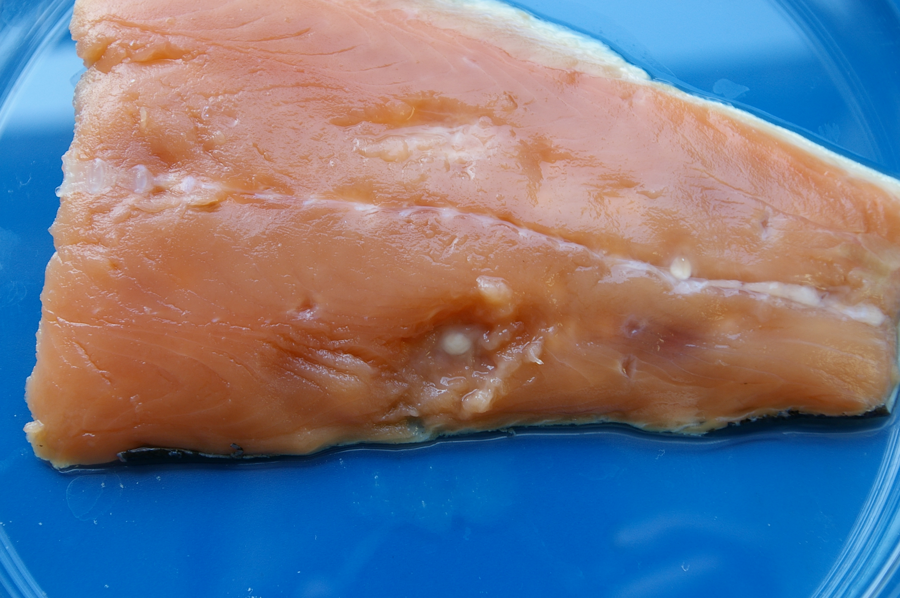
| Clinical signs | White cysts (a few mm) are observed in the trunk muscle (Figs. 1, 2). In
the frozen
fish or smoked fish, the site of infection looks like 'a milky condition' (Awakura and Kimura, 1977). |
| Parasitology | Cysts are filled with a number of spores (Fig. 3). A spore (average length
10.7 μm;
average width 8.7 μm;
average thickness 6.1μm) is oval and has 2 slightly unequal polar capsules (larger one: average
length 4.2 μm; average width 2.5 μm, smaller one: 3.8 μm; 2.2 μm) and 2 caudal appendages
(longer one: average length 34.8 μm, shorter one: 28.9 μm). The life cycle is unknown. The alternate host is
probably involved in the life cycle. |
| Pathology | The parasite was encapsulated by the host’s connective tissues and formed the cysts. |
| Health hazard | Since this parasite is not infectious to human, it is harmless in food hygiene. |
| Diagnosis | Check the spores by the wet-mount of cysts or degenerated muscle tissue. Sample should be smeared and stained by Giemsa. |
| Other information | Some authors suggested that Henneguya salminicola is synonym of Henneguya zschokkei (Lom and Dykova, 1992). |
| References | Awakura T. and T.
Kimura (1977): On the milky condition in smoked coho salmon (Oncorhynchus kisutch) caused by
myxosporidian parasite. Fish Pathol.,
12, 179-184.
Lom, J. and I. Dykova (1992): Protozoan parasites of fishes. Elsevier, New York, 315p. |

(Photos by Jun Araki)
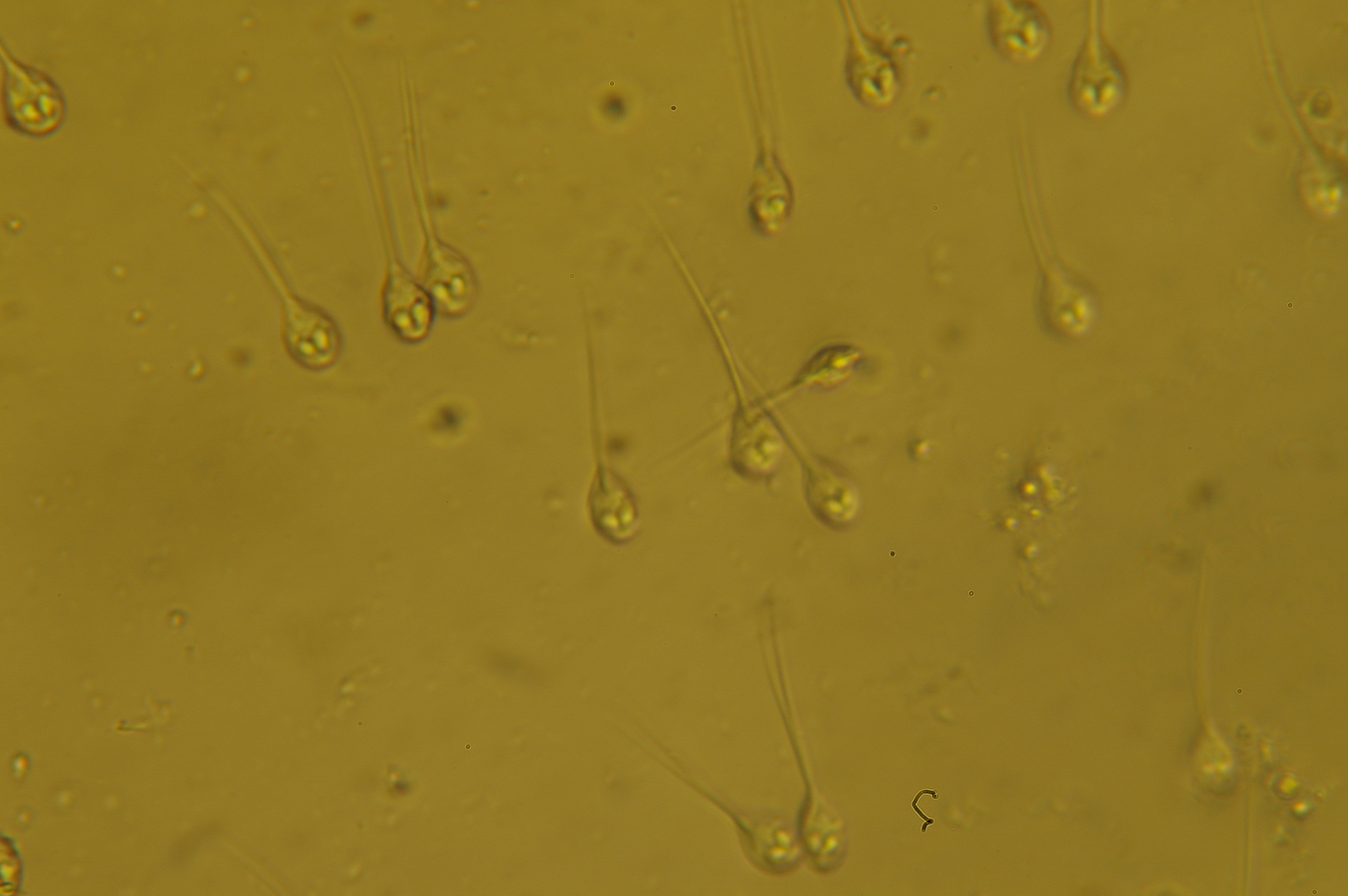
Fig. 2. Lesions caused by H. salminicola
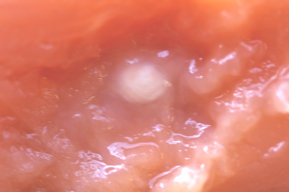
Fig. 1. Trunk muscle of pink salmon infected with Henneguya.