| Parasite | Henneguya pagri |
|---|---|
| Taxonomy | Myxozoa, Myxosporea, Bivalvulida |
| Host | Red sea bream (Pagrus major) |
| Disease name | Cardiac henneguyosis |
| Infection site | Heart |
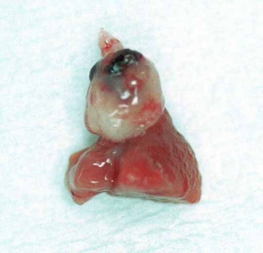
| Clinical sign | Diseased fish exhibits anaemia of the gills and the internal organs, haemorrhage in the pericardial cavity (Fig. 1) and enlargement of the bulbus arteriosus (Fig. 2). |
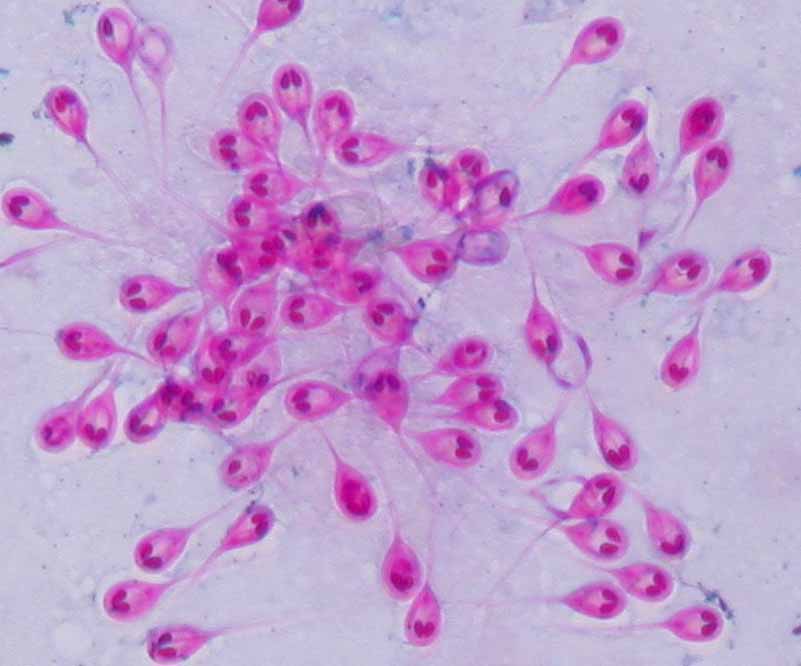
| Parasitology | A number of spores fully occludes the capillary of the gill filament (Fig. 3).
A spore (length 9.9-11.9 (average 10.5) mm; width 6.4-8.4 (7.5) mm; thickness 5.4-6.4 (5.9) mm) is ovoid and has 2 polar capsules (length 2.5-4.0 (3.1) mm; width 1.5-2.0 (1.6) mm ) and 2 caudal appendages (24.8-34.7 (29.6) mm). The life cycle is unknown. |
| Pathology | After maturaion of parasite in the bulbus arteriosus, phagocytized spores are transfered into the gill filament, causing occlusion of the capillary, congestion, lamellar fusion and destruction of the gill epithelia (Fig. 4). Severe foci are also observed in the heart, e.g. the destruction of the myocardium (Yokoyama et al., 2005). |
| Health hazard | Since this parasite is not infectious to human, it is harmless in food hygiene. |
| Diagnosis | Check the spores by wet-mount of squashed gill filaments. Henneguya pagri can be distinguished from Henneguya lateorabracis by the length (shorter in H. pagri) and shape (a whip-like extension in H. pagri) of the caudal appendages. Sample should be smeared and stained by Ziehl-Neelsen. |
| Other information | This disease occurs in 0-year fish on August to November. Though no control measures are available, diseased fish may recover after spore discharge. |
| References | Yokoyama, H., N. Itoh and S. Tanaka (2005): Henneguya pagri n. sp. (Myxozoa: Myxosporea) causing cardiac henneguyosis in red sea bream, Pagrus major (Temminck &Schlegel). J. Fish Dis., 28, 479-487. |
Fig. 1. Diseased red sea bream showing the anaemia of gill and the haemorrhages in the pericardial cavity
Fig. 2. Enlargement of the bulbus arteriosus caused by H. pagri.
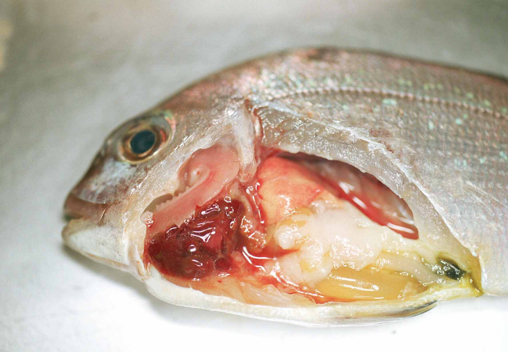
Fig. 4. Lamellar fusion and occlusion of the capillary are evident.
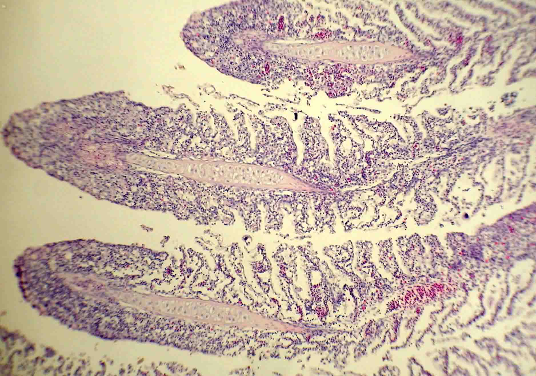
Fig. 3. Spores of H. pagri. Ziehl-Neelsen stain.