| Parasite | Heterosporis anguillarum |
|---|---|
| Taxonomy | Microspora, Microsporea |
| Hosts | Japanese eel (Anguilla japonica) |
| Disease name | Beko disease ('beko' means uneven body surface.) |
| Infection site | Trunk muscle, muscle in the stomach (ocassionally) |
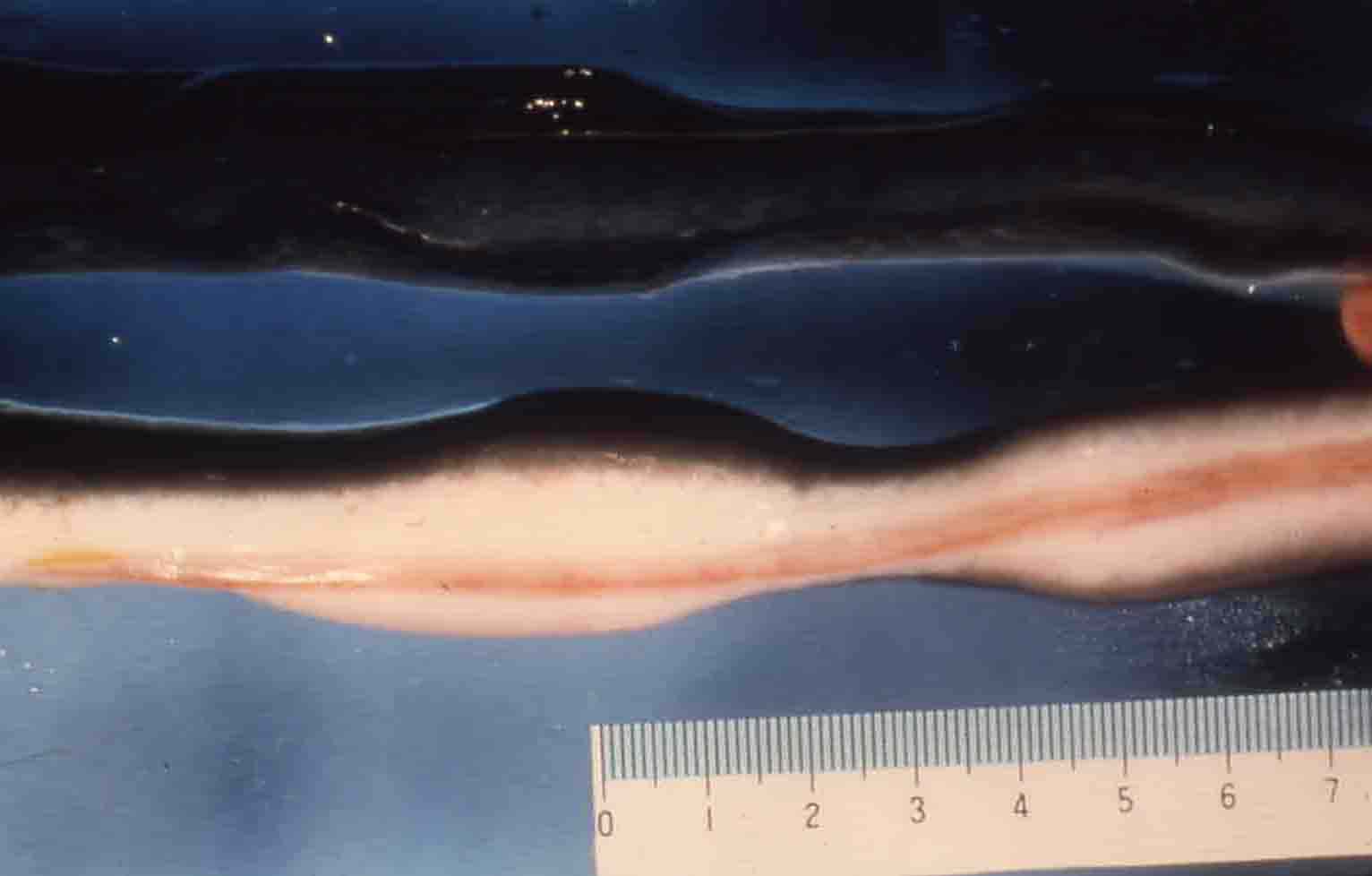
| Clinical signs | Uneven concave body surface is observed (Fig. 1). Diseased eel exhibits the whitish muscle lesion liquefied by the proteolytic enzymes of the parasite. |
| Parasitology | Spores are produced inside the cysts formed in the myocytes (Fig. 2). Heterosporis anguillarum formed two types of spores (macrospore: length 6.7-9.0 mm; width 3.3-5.3 mm, microspore: length 2.8-5.0 mm; width 2.0-2.9 mm). |
| Pathology | Cysts (up to 200 mm in diameter) are formed in the muscle tissue (Fig. 3). No obvious host reactions and lesions are observed during the development of the parasite. However, after maturation of the parasite, liquefaction of the neighboring muscle tissues occurs, possibly caused by the proteolytic enzymes from the ruptured cysts. Subsequently, infiltration of phagocytic cells and other lymphocytes are also observed. The lesions are recovered finally (Ogawa, 2004). |
| Health hazard | Since this parasite is not infectious to human, it is harmless in food hygiene. |
| Diagnosis | Check the spores by wet-mount of the lesions. However, the spores may not be detected when the fish is recovering (even when they show a typical disease sign). The sample should be smeared and stained by Uvitex 2B followed by a fluorescent microscopic observation. The stained spores emit blue fluorescence under UV radiation. |
| Other information | This disease was reported from Japan, Taiwan and Korea (Joh et al., 2007). Heterosporis anguillarum infects orally or through the skin. Spores possibly reinfect inside the fish (autoinfection), but this hypothesis has not been elucidated. Development of the parasite is influenced by the water temperature; higher water temperature induces faster development of the parasite between 20-30 C., the cysts were formed in 30 days at 25 C., and the parasite doesn’t develop (viz. the eels doesn’t develop the disease) below 15 C (Ogawa, 2004). It is recommended to prevent the cannibalism and to remove the diseased fish. Though fumagillin, an antibiotic, is partially effective, this can’t be used practically (Kano et al. 1982). |
| References | .Joh, S. J., Y. K.,
Kwon, M. C., Kim, M. J., Kim, H. M., Kwon, J. W. Park, J. H. Kwon and J. H.
Kim. (2007): Heterosporis anguillarum
infections in farm cultured eels (Anguilla
japonica) in Korea. J. Vet. Sci., 8, 147-149. Kano, T., T. Okauchi and H. Fukui (1982): Studies on Pleistophora infection in eel, Anguilla japonica. II. Preliminary test for application of fumagillin. Fish Pathol., 17, 107-114. Ogawa, K. (2004): Protozoan diseases. Infectious and parasitic diseases of fish and shellfish. (Wakabayashi, H. and K. Muroga), Koseisha koseikaku, pp.285-338. (In Japanese) |
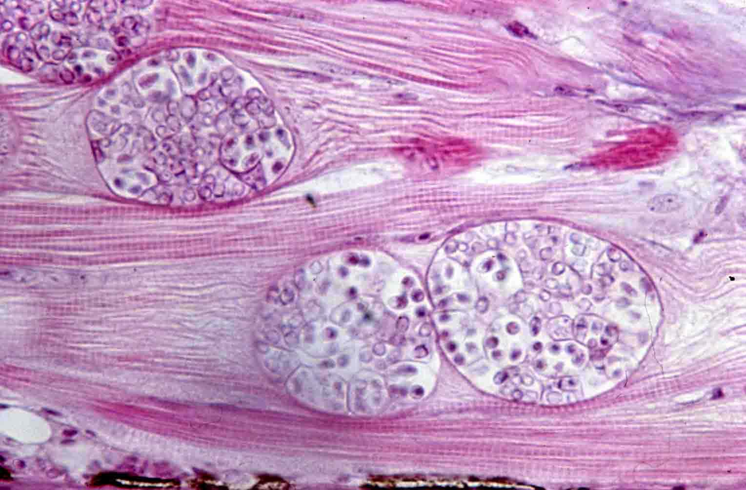
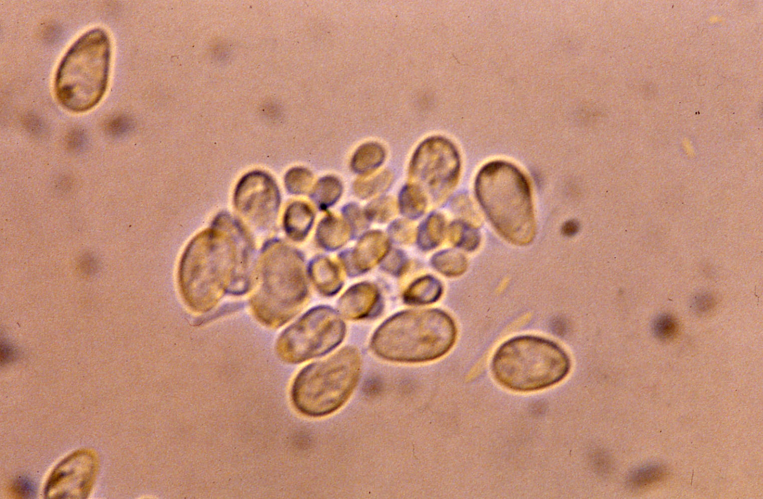
Fig. 3. Cysts developing in the myocytes of Japanese eel.
Fig. 2. Fresh spores of H. anguillarum.
Fig. 1. External sign of diseased fish.

(Photos by H. Oka)