| Parasite | Ichthyophthirius multifiliis |
|---|---|
| Taxonomy | Ciliophora, Oligohymenophorea |
| Hosts | Many warm water fishes living in freshwater. |
| Disease name | White spot disease |
| Infection site | Subepithelial tissues of the skin, fin and gill. |
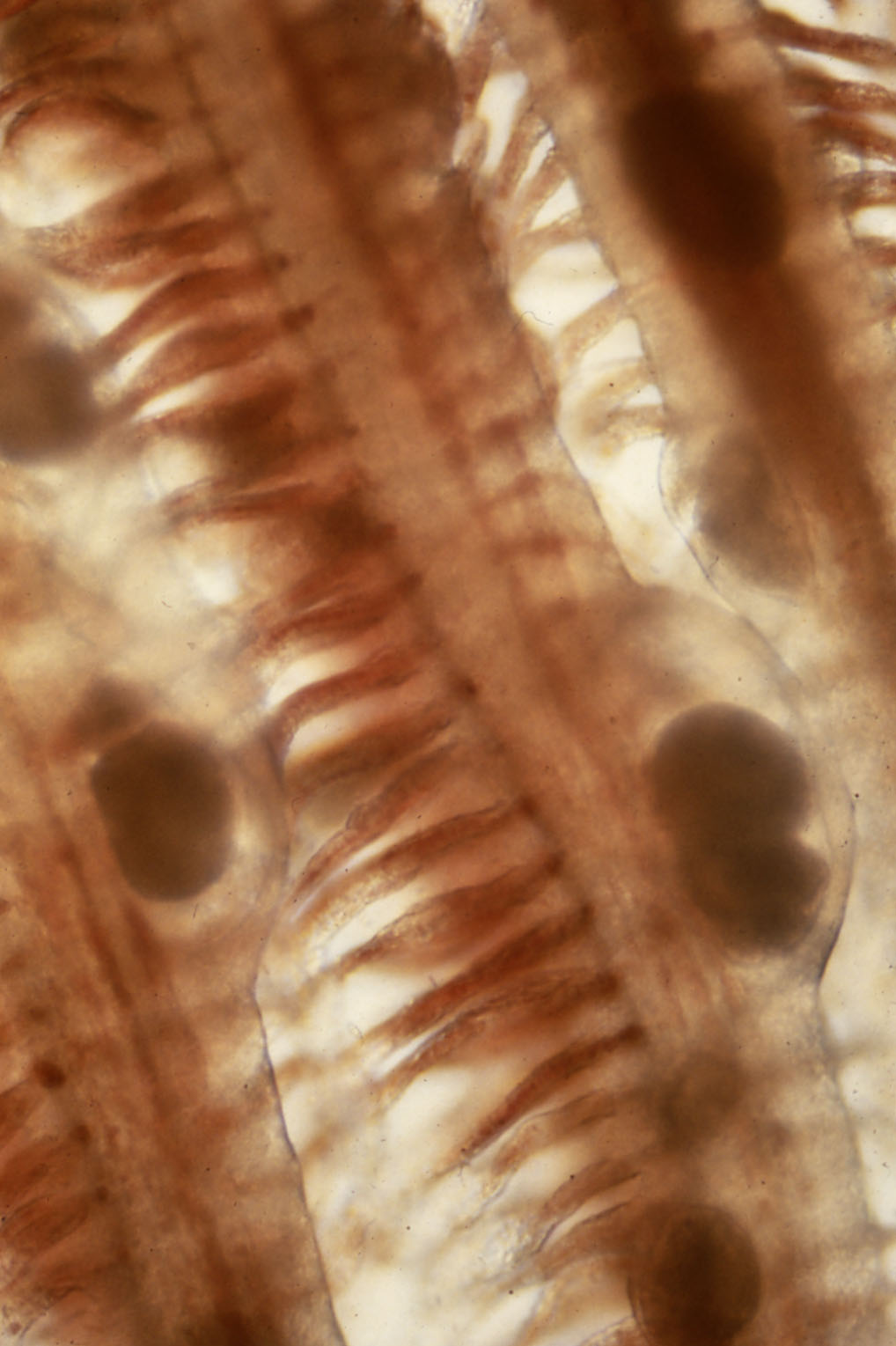
| Clinical signs | White spots appear on the entire body. Heavily infected fish exhibit mucous secretion and abnormal swimming. Rotating parasites are observed in the epithelial tissues of the skin, fin and gill under a stereomicroscope (Fig. 1). |
| Parasitology | Parasitic stage in the skin or gills of fish is the trophont. It is oval to round (0.5-1.0 mm in diameter) and contains a horseshoe-shaped macronucleus. The trophont escapes from the host in about 1 week and encysts on the bottom of water. Within its cyst, the tomont divides to produce the small tomite, which breaks through the cyst wall to become the theront. The theront penetrates into a host (Lom and Dykova, 1992; Ogawa, 2004). |
| Pathology | Mucous cells proliferate in the surface tissue and inflammatory infiltration occurs in heavily infected fish. The epithelial tissues of the skin and gills disintegrate associated with the detachment of the trophont, leading the fish to failure of the osmoregulation and respiration. |
| Health hazard | Since this parasite is not infectious to human, it is harmless in food hygiene. |
| Diagnosis | Confirm the cilia and a horseshoe-shaped macronucleus of the parasite by microscopy. |
| Other information | This disease is well-studied because it is one of the most important parasitic diseases in cultured, ornamental and aquarium fishes. Control measures can be referred elsewhere (Lom and Dykova, 1992; Ogawa, 2004). |
| References | Lom, J and I. Dykova (1992) Protozoan Parasites of Fishes, Developments
in Aquaculture and Fisheries Science, 26, Elsevier, pp. 315. Ogawa, K. (2004): Protozoan diseases. Infectious and parasitic diseases of fish and shellfish. (ed. by Wakabayashi, H. and K. Muroga), Koseisha koseikaku, pp.285-338. (In Japanese) |

(Photo by S. Egusa)
Fig. 1. The gill of Japanese eel infected with
Ichthyophthirius.