
| Parasite | Kudoa musculoliquefaciens |
|---|---|
| Taxonomy | Myxozoa, Myxosporea, Multivalvulida |
| Hosts | Swordfish (Xiphias gladius), Okhotk atka mackerel (Pleurogrammus azonus) |
| Disease name | Post-mortem myoliquefaction |
| Infection site | Trunk muscle |
| Clinical sign | The muscle tissue is liquefied after the catch (Fig. 1: Okhotk atka mackerel). Infection is not fatal. |
| Parasitology | This parasite develops inside the myofibrils and forms many spores. A spore (length 5.32〜7.28 μm; width 7.42〜9.94 μm) is quadrate-rotundate in apical view (Fig. 2) and has 4 polar capsules (1.68〜2.80 μm ). The life cycle is unknown. |
| Pathology | The myofibrils are disintegrated and the neighboring tissues are liquefied by the proteolytic enzymes derived from the parasite. |
| Health hazard | Since this parasite is not infectious to human, it is harmless in food hygiene. There is no report that the liquefied fish meat is toxic. |
| Diagnosis | Check the spores by wet-mount of the squashed muscle tissue. When the myoliquefactions progress, the detection of spores is sometimes difficult. Sample should be smeared and stained by Giemsa or Diff-Quik. |
| Other information | In summer of 1953, this parasite was found from swordfish caught at Kesennuma, northeastern Japan, and described as Chloromyxum musculoliquefaciens. Later, it was transferred to the genus Kudoa (Egusa, 1986; Matsumoto, 1954). Recently, K. musculoliquefaciens was found in okhotk atka mackerel from Bering Sea exhibiting the post-mortem myoliquefaction. However, molecular analysis might be necessary for confirmative diagnosis. |
| References | Egusa, S. (1986): The order Multivalvulida
Shulman, 1959 (Myxozoa; Myxosporea): a review. Fish Pathol., 21,
261-274. Matsumoto, K. (1954): On the two new myxosporidia, Chloromyxum musculoliquefaciens sp. nov. and Neochloromyxum cruciformum gen. et ap. Nov., from the jellied muscle of swordfish, Xiphias gladius Linne, and common Japanese sea-bass, Lateolabrax japonicus (Temminck et Schlegel). Bull. Jap. Soc. Sci. Fish., 20, 469-478, Pl. 1. |
Fig. 1. Okhotk atka mackerel exhibiting muscle liquefaction.
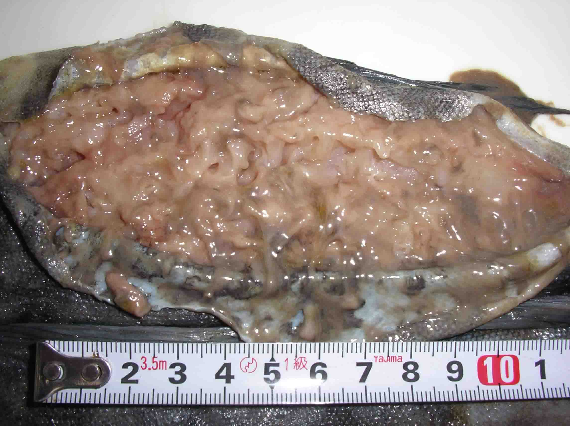
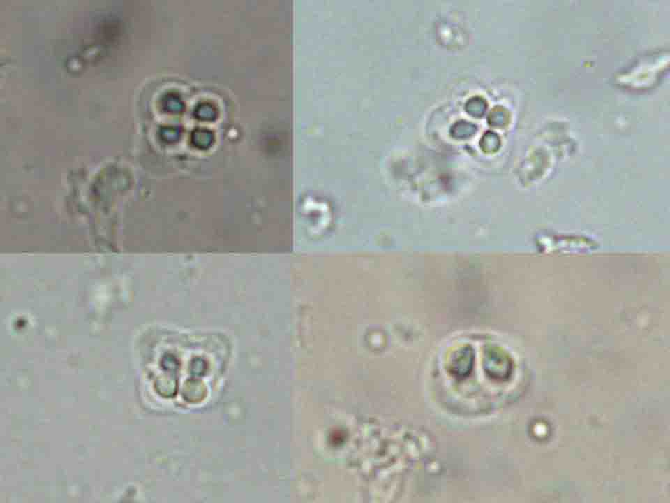
Fig. 2. Fresh spores of K. musculoliquefaciens from the
Okhotk atka mackerel.