| Parasite | Kudoa trachuri |
|---|---|
| Taxonomy | Myxozoa, Myxosporea, Multivalvulida |
| Hosts | Japanese jack mackerel (Trachurus japonicus |
| Infection site | Trunk muscle |
| Clinical sign | White fusiform cysts up to 7 mm are formed. A myoliquefaction does not occur. This disease is nonfatal to host fish. |
| Parasitology | Many spores are produced inside the cysts (Fig. 3). A spore (length 5.3〜6.5 μm; width; 8.5〜9.8 μm) is quadrate-rotundate in apical view. The life cycle is unknown. |
| Pathology | No report |
| Health hazard | Since this parasite is not infectious to human, it is harmless in food hygiene |
| Diagnosis | Check the spores by wet-mount of cysts (Egusa, 1986). Sample should be smeared and stained by Giemsa or Diff-Quik. |
| Other information | In these days, K. trachuri was sometimes found from wild Japanese jack mackerel in East China Sea. |
| References | Egusa,
S. (1986): The order Multivalvulida Shulman, 1959 (Myxozoa; Myxosporea): a
review. Fish Pathol., 21, 261-274. (In Japanese) Matsukane, Y., H. Sato, S. Tanaka, Y. Kamata and Y. Sugita-Konishi (2011): Kudoa iwatai and two novel Kudoa spp., K. trachuri n. sp. and K. thunni n. sp. (Myxosporea: Multivalvulida), from daily consumed marine fish in western Japan. Parasitol. Res., 108, 913-926. |
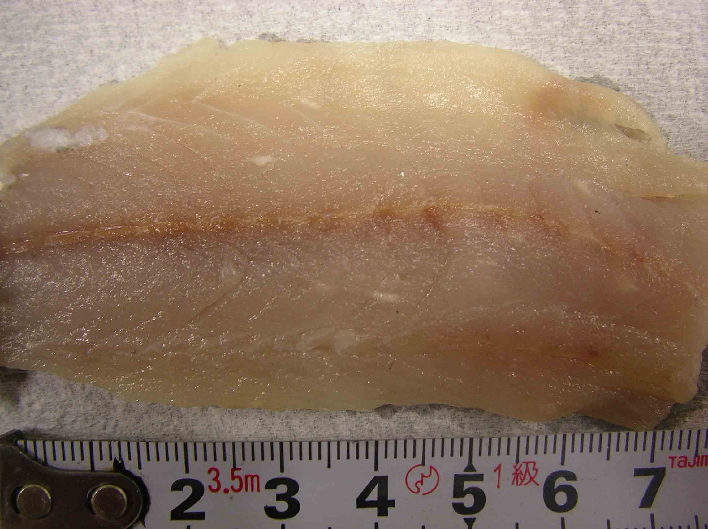
Fig. 1. A fusiform cyst of Kudoa trachuri in trunk muscle of
Japanese jack mackerel.
Fig. 3. Fresh spores of Kudoa trachuri
(Photo by R. Kuranaga(1))

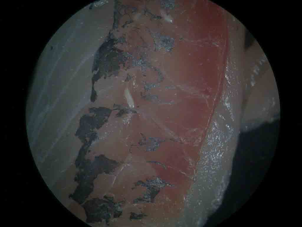
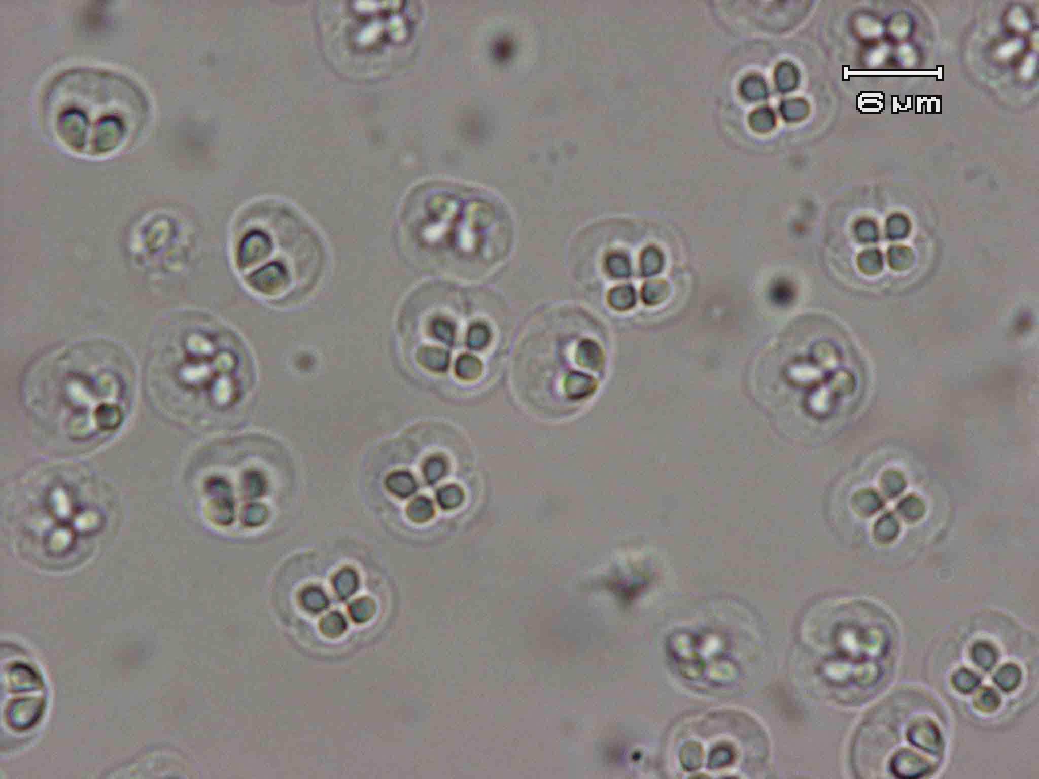
Fig. 2. Several white cysts of Kudoa trachuri.