| Parasite | Kudoa sp. |
|---|---|
| Taxonomy | Myxozoa, Myxosporea, Multivalvulida |
| Hosts | Bar-tailed flathead (Platycephalus sp.) |
| Infection site | Trunk muscle |
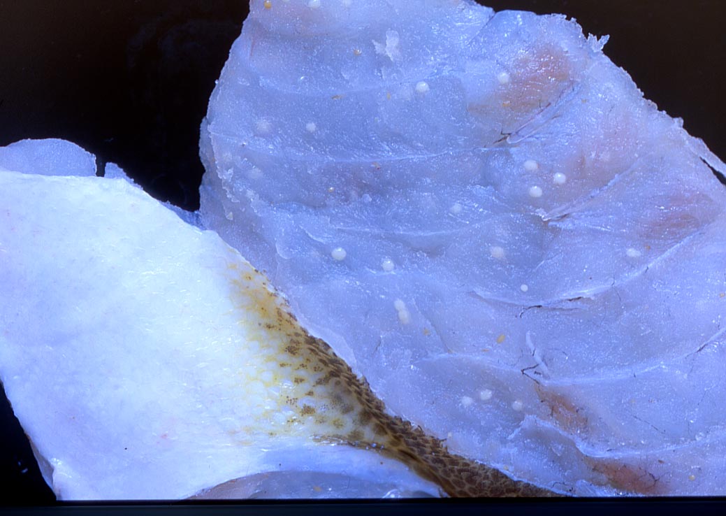
| Clinical sign | Many white spherical cysts (1-2 mm) are usually observed (Fig. 1), while brown elliptical cysts (up to 0.5 mm) are occasionally found. |
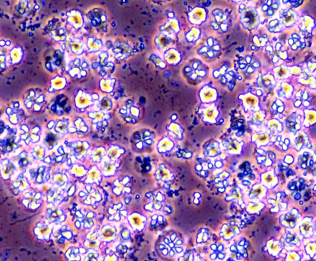
| Parasitology | Many spores are produced inside cysts (Fig. 2). A spore (average length 8.5 mm; average width 10.5 mm) has 4 polar capsules. The life cycle is unknown. |
| Health hazard | Since this parasite is not infectious to human, it is harmless in food hygiene. |
| Diagnosis | Check the spores by wet-mount of cysts. Sample should be smeared and stained by Giemsa or Diff-Quik. |
| Other information | This parasite seems to be closely related with Kudoa iwatai, a parasite of red sea bream based on the morphology of cysts and spores (Momoyama and Tensha, 2006), but has not been identified yet. |
| References | Momoyama, K. and K. Tensha (2006): Ugly-looking parasitic infections and
other abnormalities of wild fish and shellfish caught in the coastal or
inland waters around or in Yamaguchi Prefecture. Bull. Yamaguchi Pref. Fish. Res. Ctr.,
4, 143-161. (In Japanese) |
(Photos by K. Momoyama)
Fig. 2. Fresh spores of Kudoa sp.
Fig. 1. Kudoa cysts in the muscle of bar-tailed flathead.
