
| Parasite | Kudoa shiomitsui |
|---|---|
| Taxonomy | Myxozoa, Myxosporea, Multivalvulida |
| Hosts | Tiger puffer (Takifugu rubripes), Greater amberjack (Seriola dumerili), Indean perch (Apogon lineatus), Japanese flounder (Paralichthys olivaceus) |
| Infection site | Heart (pericardial cavity) |
| Clinical sign |
White spherical cysts (ca. 1mm diameter) are observed at the surface of heart and pericardial cavity (Fig. 1). In some fish species, the vegetative stages are observed in the gill filaments (Fig. 2). |
| Parasitology | Many spores are produced inside the cysts (Fig. 3). A quadrate spore (width 9-10 μm) has 4 polar capsules. The life cycle is unknown. |
| Pathology | It has been considered that this parasite has a low pathogenicity because the cysts are encapsulated by the host tissues and adhered lightly to the surface of the heart. However, it is likely that a swimming ability is impaired in case of heavy infection. |
| Health hazard | Since this parasite is not infectious to human, it is harmless in food hygiene. |
| Diagnosis | Check the spores by wet-mount of cysts. K. shiomitusi can be distinguished from Kudoa pericardialis, parasitizing the pericardial cavity of yellowtail, by the spore width (6-7 μm for K. pericardialis). Sample should be smeared and stained by Giemsa or Diff-Quik. |
| Other information | Fishes reared by unfiltered sea water could be infected with the parasite even in a land-based tank. A causal relation between the parasite and deteriorated swimming ability should be further studied. |
| References | Egusa, S. and T. Shiomitsu (1983): Two new species of the genus Kudoa (Myxosporea: Multivalvulida) from marine cultured fishes in Japan. Fish Pathol., 18,163-171. (In Japanese) |
Fig. 1. Numerous cysts of K. shiomitsui in the pericardial cavity
of tiger puffer.
(Photo by K. Ogawa (1))
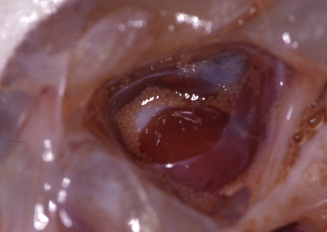
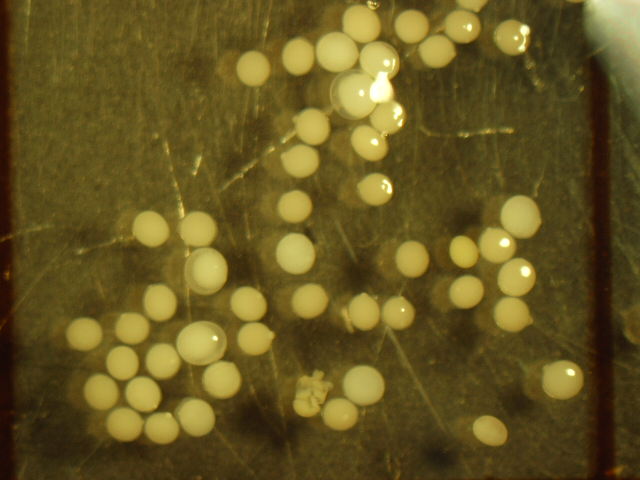
Fig. 2. Cysts of K. shiomitsui collected from tiger puffer.
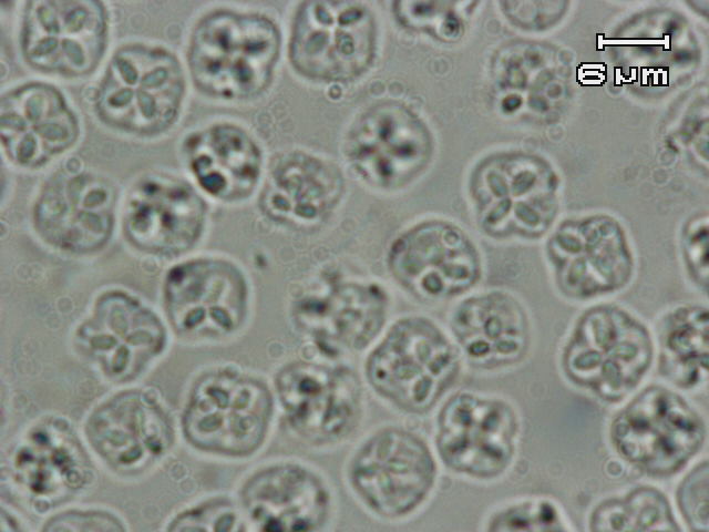
Fig. 3. Fresh spores of K. shiomitsui