| Parasite | Kudoa yasunagai |
|---|---|
| Taxonomy | Myxozoa, Myxosporea, Multivalvulida |
| Hosts | Yellowtail (Seriola quinqueradiata), Red sea bream (Pagrus major), Tiger puffer (Takifugu rubripes), Japanese flounder (Paralichthys olivaceus), Japanese sea bass, (Lateolabrax japonicus), Japanese striped knifejaw (Oplegnathus fasciatus), Pacific bluefin tuna(Thunnus orientalis)、Striped eel catfish (Plotosus lineatus) |
| Infection site | Brain |
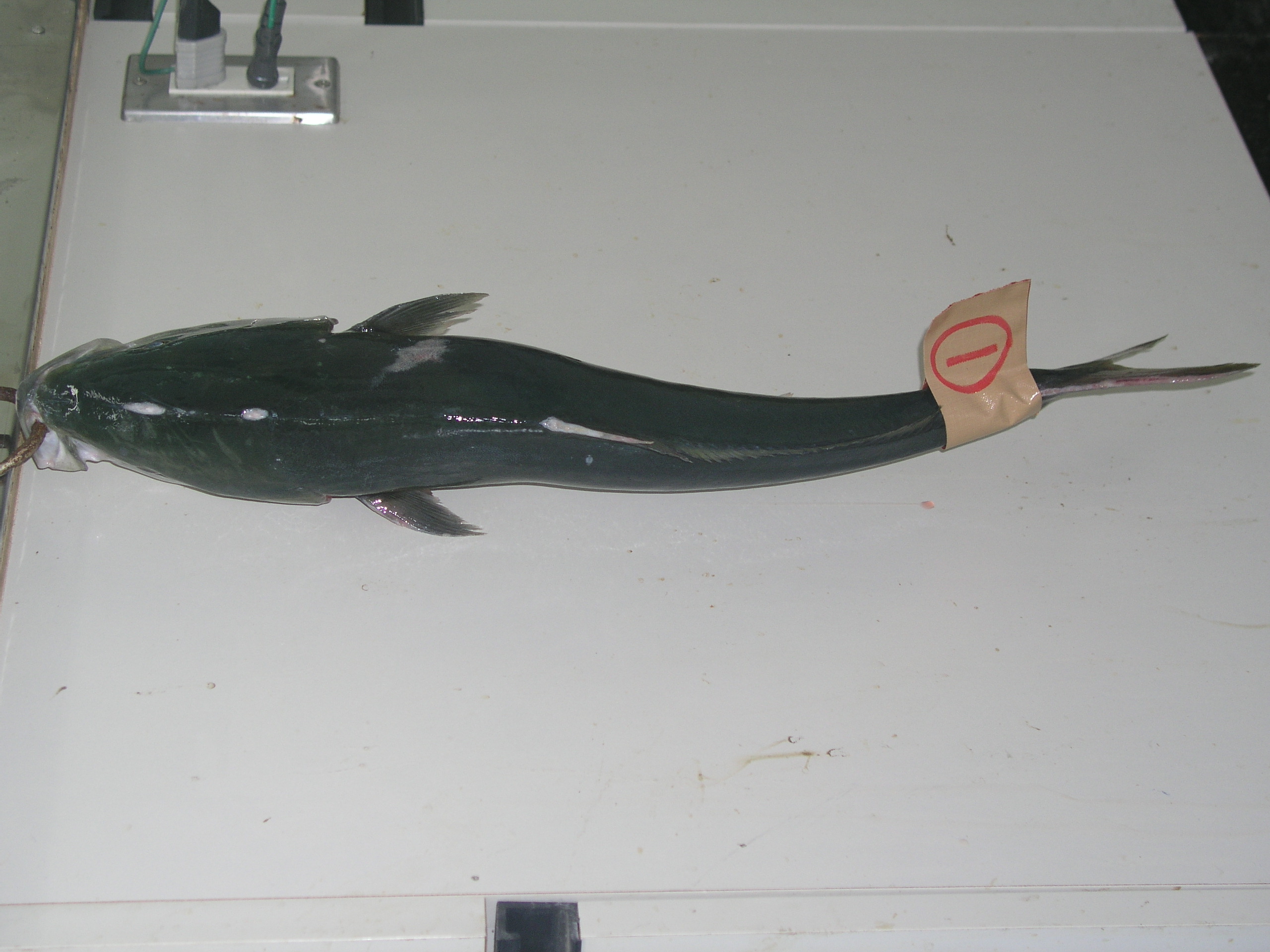
| Clinical signs | White small spherical cysts are observed around the brain (Fig. 1). Diseased fishshows the abnormal behavior and the deformation. |
| Parasitology | Many spores were produced inside the cysts (Fig. 2). A spore (average length 6.2 mm; average width 11.7 mm; average thickness 8.3 mm) has 6-8 (in general, 7) polar capsules (average length 3.6 mm; average width 2.4 mm). |
| Pathology | Cysts were observed in and at the surface of the brain, medulla oblongata and the anterior part of spinal cord. A causal relationship between the abnormal swimming and the infection site is unknown. |
| Health hazard | Since this parasite is not infectious to human, it is harmless in food hygiene. |
| Diagnosis | Check the spores by wet-mount of cysts. Kudoa yasunagai can be distinguished from Myxobolus acanthogobii, parasitizing the brain of marine fishes, by the number of polar capsules (2 for M. acanthogobii). Sample should be smeared and stained by Giemsa or Diff-Quik. |
| Other information | This parasite was firstly reported from cultured Japanese sea bass and Japanese striped knifejaw Oplegnathus fasciatus at Nagasaki prefecture in 1980 (Yasunaga et al., 1981). These fish swam abnormally and died. Because of 7 spore valves and 7 polar capsules, this parasite was described as the new family (Septemcapsulidae), genus and species (Septemcapsula yasunagai) (Hsieh and Chen, 1984). However, this species was transferred to the genus Kudoa as a result of the molecular phylogenic analysis (Whipps et al., 2004). There are no effective methods to prevent this disease. |
| References | Arthur, J. R. and S. Lumanlan-Mayo (1997): Checklist of the parasites of fishes of the Philippines. FAO Fisheries Technical Paper, 369, 102 p. FAO, Rome. Hsieh, S. R. and C. L. Chen (1984): Septemcapsula yasunagai gen. et sp. nov., representative of a new family of the class Myxosporea. Acta Zootaxon. Sin., 9, 225-227. Yasunaga, N., K. Hatai, S. Ogawa and S. Yasumoto (1981): An unknown Myxozoa found in brain of cultured sea bass, Lateolabrax japonicus and cultured Japanese striped knifejaw, Oplegnathus fasciatus. Fish Pathol., 16, 51-54. (In Japanese) Whipps, C. M., G. Grossl, R. D. Adlard, H. Yokoyama, M. S. Bryant, B. L. Munday and M. L. Kent (2004): Phylogeny of the Multivalvulidae (Myxozoa: Myxosporea) based on comparative ribosomal DNA sequence analysis. J. Parasitol., 90, 618-622. Zhang, J.Y., F. Meng, H. Yokoyama, J. Miyahara, I. Takami and K. Ogawa (2010): Myxosporean and microsporidian infections in cultured Pacific bluefin tuna Thunnus orientalis in Japan. Fish Sci., 76, 981-990.. |

Fig. 3. Fresh spores of Kudoa yasunagai
(Photo by H. Matsuo (1))
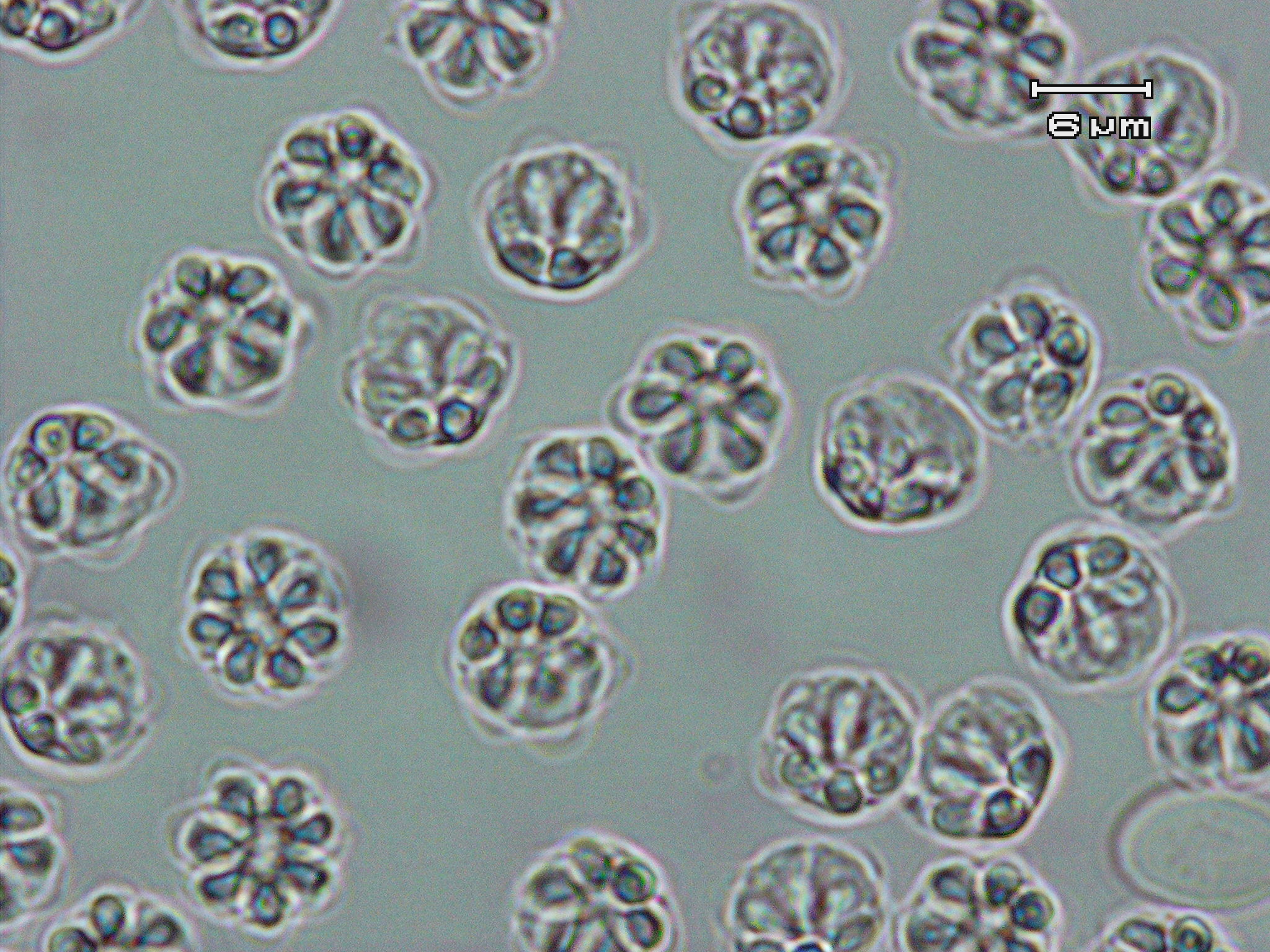
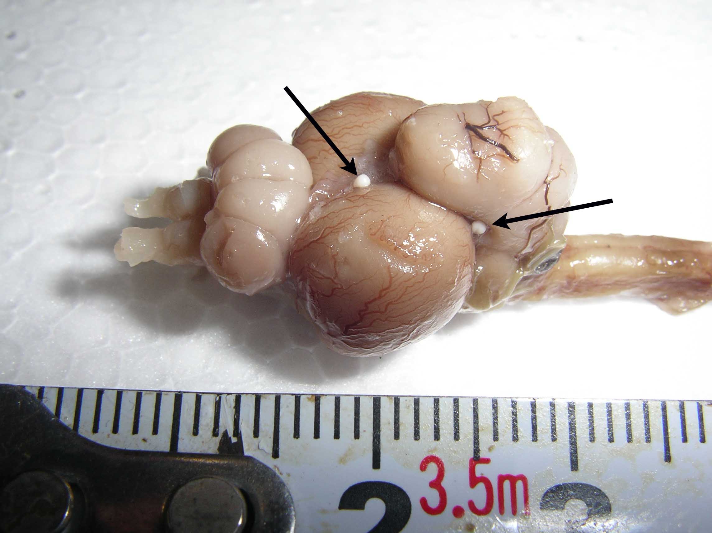
Fig. 2. Cysts (arrows) of Kudoa yasunagai on the brain of yellowtail.
Fig. 1. Deformed yellowtail infected with K. yasunagai.