| Parasite | Lepeophthirus salmonis (Sea lice) |
|---|---|
| Taxonomy | Arthropoda, Crustacea, Copepoda, Siphonostomatoida |
| Hosts | Salmonids including Atlantic salmon (Salmo salar), chum salmon (Oncorhynchus keta) and pink salmon (O. gorbuscha). |
| Infection site | Skin |
| Clinical signs | Lesions caused by sea louse are observed in the skin (mainly basal region of the anal fin and head) (Fig. 1). |
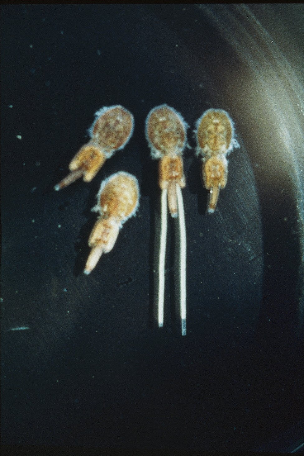
| Parasitology | Lepeophtheirus salmonis is an exoparasitic crustacean and ca. 1 cm in size. A female possesses a pair of egg sacs in the posterior end of the body (Fig. 2). The number of larval stages is 2 nauplius, 1 copepodid, 4 chalimus and 2 preadult. The copepodid settles on the host’s skin (Johnson and Albright, 1991). The settled parasite feeds on host’s epidermis and mucus. |
| Pathology | The sites of heavy infection show abrasion and haemorrhage. Such lesions sometimes expose the muscle. The disease is often accompanied with the secondary infection of bacteria. |
| Health hazard | Since this parasite is not infectious to human, it is harmless in food hygiene. |
| Diagnosis | Morphologically observe the adults. Lepeophtheirus can be differentiated from Caligus because Lepeophtheirus lacks a pair of prominent lunules in the anterior end. |
| Other information | This disease is well studied because it is one of the most important diseases of Atlantic salmon culture in Europe. It is recommended to practice the combination of chemotherapeutic treatments, managements of culture program (e.g., fallowing) and use of the cleaner fish (Pike and Wadsworth, 2000). Sea lice is not so harmful in Japanese aquaculture, though the light infection occurs in cultured coho salmon in Tohoku area. |
| References | Johnson, S. C. and L. J.
Albright (1991): The developmental stages of Lepeophtheirus salmonis (Kroyer, 1837) (Copepoda: Caligidae). Can J. Zool., 69, 929-950.
Pike, A. W. and S. L. Wadsworth (2000): Sealice on salmonids: Their biology and control. Adv. Parasitol., 44, 233-337. |
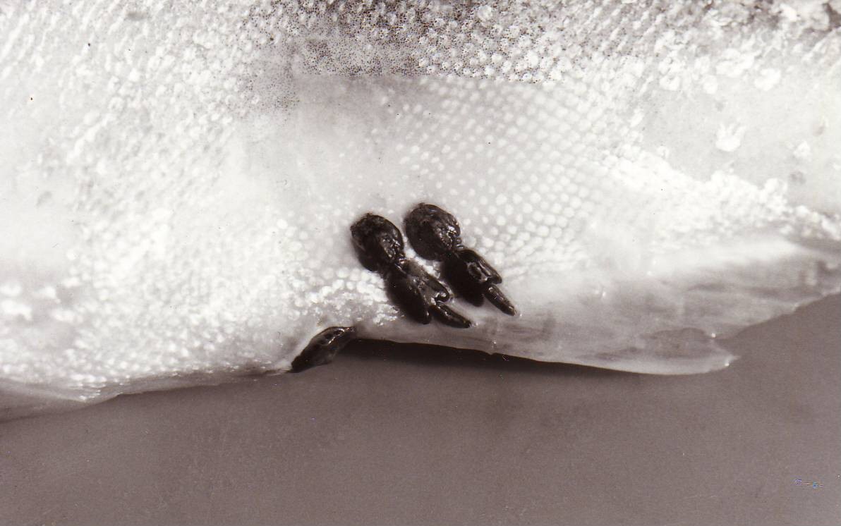

Fig. 1. Sea louse on the basal part of the anal fin.
Fig. 2. Three males and one female (with egg sacs) of
sea lice
(Photos by K. Nagasawa (1) and T. Awakura (2))