| Parasite | Myxobolus sp. |
|---|---|
| Taxonomy | Myxozoa, Myxosporea, Bivalvulida |
| Hosts | Crucian carp (Carassius auratus), Prussian carp (Carassius gibelio) |
| Infection site | Head, fin, skin |
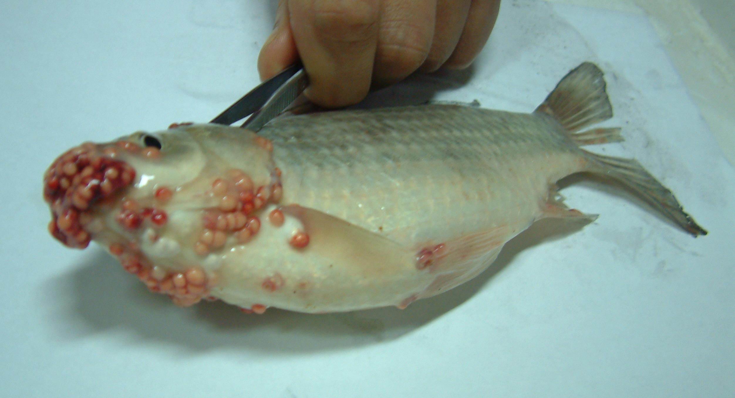
| Clinical signs | Many white cysts are observed on the head, in particular, the mouth region (Fig. 1). |
| Parasitology | A number of spores are formed inside the cyst (Fig. 2). A spore (length 9.2 (8.4-9.8) mm; width 8.2 (7.8-8.4) mm; thickness 5.5 (5.4-5.8) mm) is almost round and has 2 equal polar capsules (length 4.5 (3.6-4.8) mm; width 2.8 (2.4-3.0) mm) (Chen and Ma, 1998). The life cycle is unknown. |
| Pathology | Although this disease is not fatal to the host fish, the market value of infected fish is reduced (Lu et al., 2002). |
| Health hazard | Since this parasite is not infectious to human, it is harmless in food hygiene. |
| Diagnosis | Check the spores by wet-mount of squashed cysts. Sample should be smeared and stained by Giemsa or Diff-Quik. |
| Other information | This disease occurred some cultured crucian carp in China. It was identified as Myxobolus rotundus, and several immunological studies were conducted (Lu et al., 2002; Lu et al, 2003; Zhang et al., 2006). However, it is probably not the same species with M. rotundus due to their different gene sequences of SSU rDNA (K. Molnar, personal communication). |
| References | Chen, Q. L., and C. L. Ma (1998): Myxozoa, Myxosporea. Fauna Sinica. Beijing,
Science Press, 987 p.
Lu, Y. S., M. Li, Y. S. Wu and J. G. Wang (2002): Antigenic study of Myxobolus rotundus (Myxozoa: Myxosporea) using monoclonal antibodies. J. Fish Dis., 25, 307-310. Lu, Y. S. P. Nie and B. J. Sun (2003): Detection of Myxobolus rotundus (Myxozoa: Myxosporea) in skin mucus of crucian carp Carassius auratus auratus using monoclonal antibody. Dis. Aquat. Org., 54, 171-173. Zhang, J. Y., J. G. Wang, Y. S. Wu, M. Li, A. H. Li and X. L. Gong (2006): A combined phage display ScFv library against Myxobolus rotundus infecting crucian carp, Carassius auratus auratus (L.), in China. J. Fish Dis., 29, 1-7. |

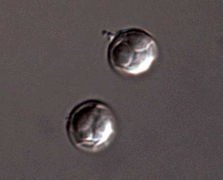
Fig. 2. Fresh spores of Myxobolus sp.
(Photos by Jin-Yong Zhang)
Fig. 1. Heavily infected crucian carp around region of the mouth.