| Parasite | Myxobolus acanthogobii |
|---|---|
| Taxonomy | Myxozoa, Myxosporea, Bivalvulida |
| Hosts | Yellowtail (Seriola quinqueradiata), Chub mackerel (Scomber japonicus), Yellowfin goby (Acanthogobius flavimanus), Red gurnard (Chelidonichthys spinosus), Gnomefish (Scombrops boops), Brown-lined puffer (Canthigaster rivulata) |
| Disease name | Myxosporean scoliosis ( for yellowtail) |
| Infection site | Brain |
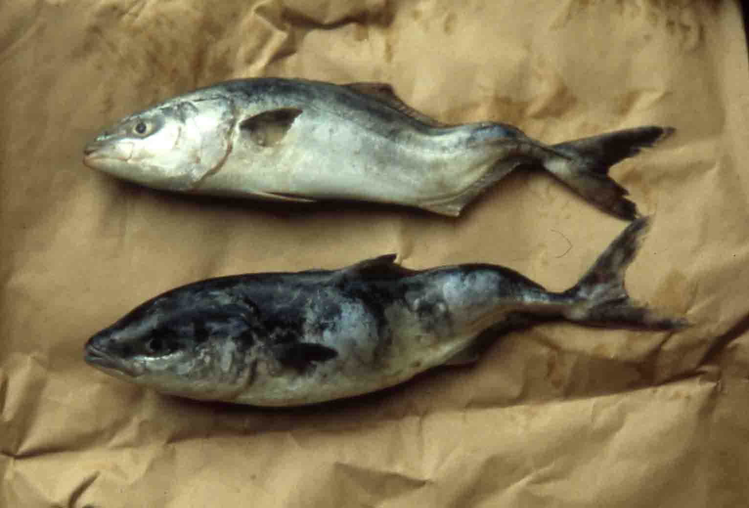
| Clinical signs | The vertebrae of infected yellowtail curves laterally (Fig. 1), while that of chub mackerel curves dorso-ventrally (Fig. 2). On the other hand, no skeletaldeformities are observed in yellowfin goby. White cysts are formed in all fish species. |
| Parasitology | Many spores are produced inside the cysts (Fig. 3). A spore (length 9.2-11.8 (average 10.6) mm; width 7.9-10.2 (9.2) mm; thickness 5.5-7.3 (6.6) mm) has 2 polar capsules (length 3.9-5.4 (4.5) mm; width 2.5-3.4 (2.8) mm). The life cycle is unknown. |
| Pathology | Cysts are encapsulated by the host tissues are observed in various parts of the brain (Fig. 4). In case of the scoliosis is yellowtail, a skeletal deformity is caused by the cysts in the forth ventricle of the brain (Maeno and Sorimachi, 1992). |
| Health hazard | Since this parasite is not infectious to human, it is harmless in food hygiene. |
| Diagnosis | Check the spores by wet-mount of cysts. Myxobolus acanthogobii can be easily distinguished from Kudoa yasunagai, parasitizing the brain of marine fishes, by the number of polar capsules (6-8 for K. yasunagai). Sample should be smeared and stained by Giemsa or Diff-Quik. The detection method by PCR was developed (Miyajima et al., 2005). |
| Other information | Several aetiological factors were suspected for the scoliosis in cultured yellowtail, but it was elucidated that this diseases is caused by infection of Myxobolus buri in the brain (Egusa, 1985). Later, M. buri was reassigned as M. acanthogobii by the molecular analysis and the morphological observation (Yokoyama et al., 2004). This parasite also causes the skeletal deformities in cultured chub mackerel (Yokoyama et al., 2005). It was reported that the consecutive feeding of artificial feed to juvenile fish could reduce the incidence of the disease (Sakaguchi et al., 1987). This result suggests that the juvenile fish is infected with this parasite by eating wild invertebrate host. |
| References | Egusa, S. (1985): Myxobolus buri sp. n. (Myxosporea:
Bivalvulida) parasitic in the brain of Seriola
quinqueradiata Temminck et Schlegel. Fish
Pathol., 19, 239-244. Maeno, Y. and M. Sorimachi (1992): Skeltal abnormalities of fishes caused by parasitism of Myxosporea. NOAA Technical Report NMFS, 111, 113-118. Miyajima, S., H. Yokoyama, Y. Fukuda, K. Okamoto and K. Ogawa (2005): A PCR method to detect Myxobolus acanthogobii (Myxozoa: Myxosporea), the causative agent of skeletal deformities of marine fishes. Fish Pathol., 40, 197-199 Sakaguchi, S., T. Hara, T. Matsusato, T. Shibahara, Y. yamagata, H. Kawai and Y. Maeno (1987): Scoliosis of cultured yellowtail caused by parasitic Myxobolus buri. Bul. Natl. Res. Inst. Aquacult., 12, 79-86. (In Japanese) Yokoyama, H., M. A. Freeman, T. Yoshinaga and K. Ogawa (2004): Myxobolus buri, the myxosporean parasite causing scoliosis of yellowtail, is synonymous with Myxobolus acanthogobii infecting the brain of the yellowfin goby. Fish. Sci., 70, 1036-1042. Yokoyama, H., M. A. Freeman, N. Itoh and Y. Fukuda (2005): Spinal curvature of cultured Japanese mackerel Scomber japonicus associated with a brain myxosporean, Myxobolus acanthogobii. Dis. Aquat. Org., 66, 1-7. |

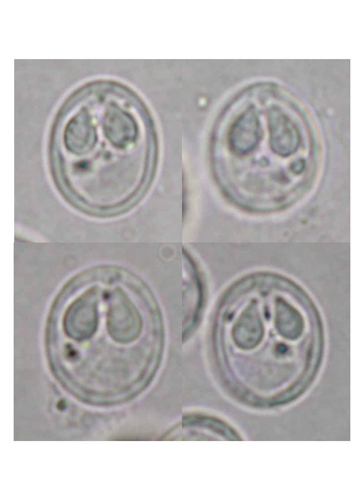
Fig. 3. Diseased chub mackerel showing dorso-lateral curvature.
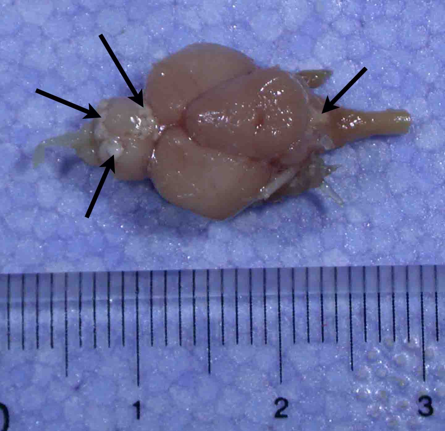
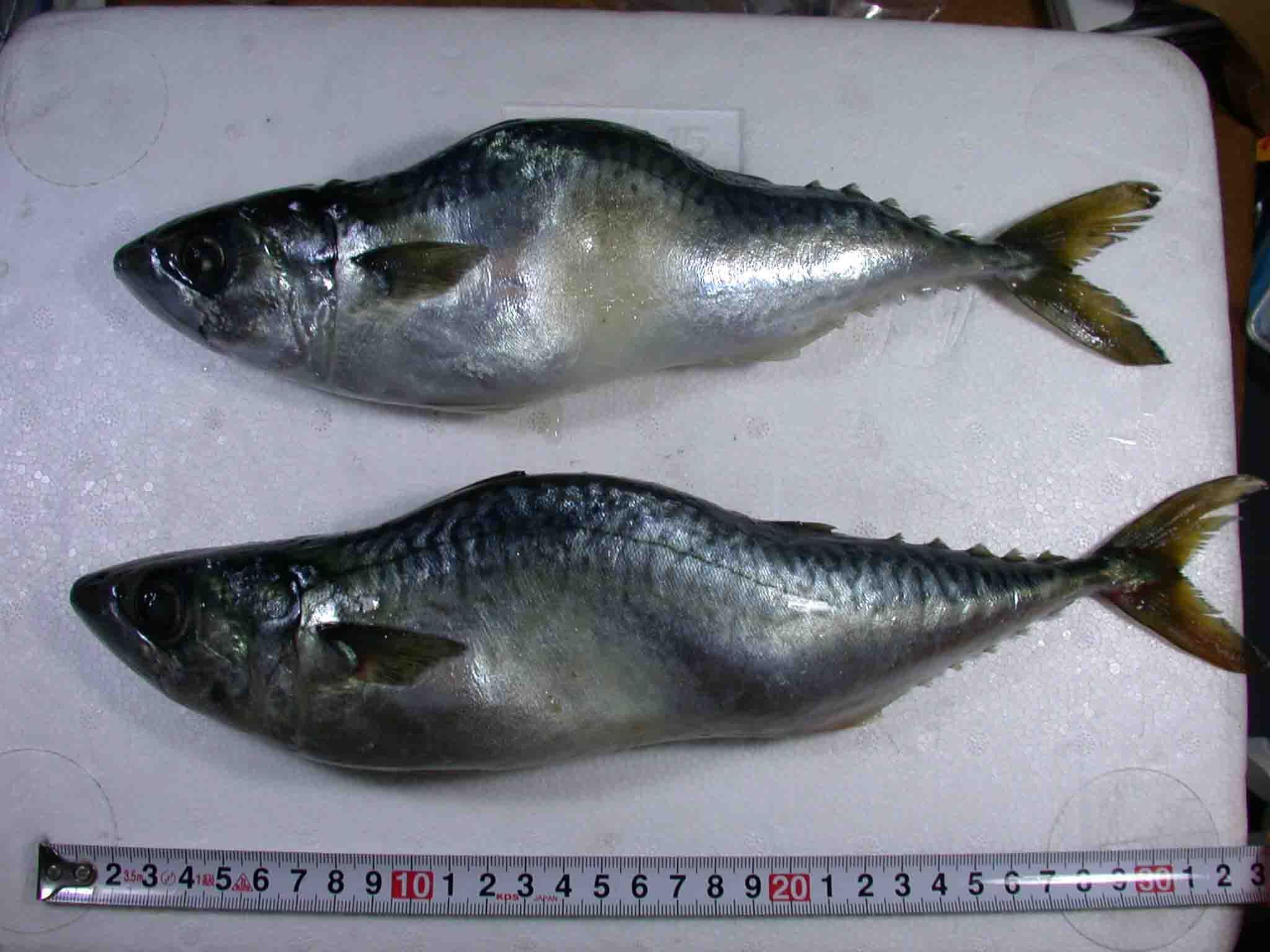
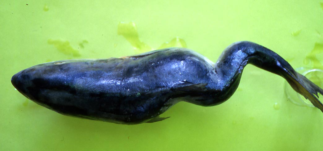
Fig. 6. Longitudinal section of the brain of chub mackerel infected with M. acanthogobii. Arrows show the cysts. OLL: olfactory lobe; OPL: optic lobe; COT: the cavity of optic tectum; C: cerebellum; FV: 4th ventricle; IL: inferrior lobe. Scale bar is 1 mm.
Fig. 5. Fresh spores of M. acanthogobii
Fig. 4. Cysts of M. acanthogobii on the brain of chub mackerel
Fig. 2. Dorsal view of diseased yellowtail.
(Photo by K. Momoyama (2))
Fig. 1. Yellowtail showing the myxosporean scoliosis.
