| Parasite | Marteilioides chungmuensis |
|---|---|
| Taxonomy | Paramyxa |
| Hosts | Pacific oyster (Crassostrea gigas), iwagaki oyster (Crassostrea nippona) |
| Infection site | Ovary |
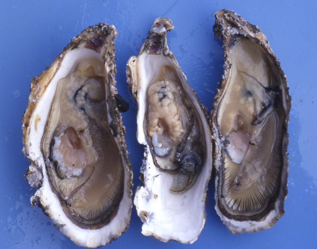
| Clinical sign | No external signs are evident. Yellowish nodular lesions (several mm to 1 cm) are observed in the ovary (Fig. 1). |
| Parasitology | Marteilioides chungmuensis is an intracellular protozoan parasite infecting the oocytes. Size of M. chungmuensis depends on the stage of development (5-25 mm in diameter) (Fig. 2). The parasite sporulates in the oocytes, and infected eggs are discharged from the host (Itoh et al., 2002). The intermediate host could be involved in the life cycle, because a free-living copepod is proven to be the intermediate host for a closely related parasite M. refringens. |
| Pathology | M. chungmuensis causes nodules in the gonad of infected Pacific oyster. These nodules give unsightly appearance to the oyster, resulting in a loss of marketability. It had been thought that pathogenicity to the host was low. However, it was reported that high mortalities of infected oyster occur in August and September. Infected oysters produce oocytes continuously in autumn, resulting in glycogen loss. As a consequence, infected oysters may suffer from nutritional stress and die (Tun et al., 2007). |
| Health hazard | Since this parasite is not infectious to human, it is harmless in food hygiene. |
| Diagnosis | The gross sign of the lumpy ovary can be used for a presumptive diagnosis because such a symptom has not been reported in other diseases of Pacific oyster. However, this symptom cannot be visually observed in the early stage and the low level of infection. Therefore, it is recommended for the definitive diagnosis to confirm the parasite in smears or histological sections. Detection methods by PCR or in situ hybridization are also developed (Itoh et al., 2003). |
| Other information | This disease used to be called as ‘abnormal egg mass.’ It occurs in fish farms of Japan and Korea, leading to substantial economic losses. Although there are no control methods for this disease, Tun et al. (2006) reported that the prevalence of infection is lower in Pacific oyster cultured in intertidal conditions. |
References |
Itoh, N., T. Oda, K.
Ogawa and H. Wakabayashi (2002): Identification and development of paramyxean
ovarian parasite in the Pacific oyster Crassostrea
gigas. Fish Pathol., 37, 23-28. Itoh, N., T. Oda, T. Yoshinaga and K. Ogawa (2003): DNA probes for detection of Marteilioides chungmuensis from the ovary of Pacific oyster Crassostrea gigas. Fish Pathol., 38, 163-169. Tun, K. L., N. Itoh, H. Komiyama, N. Ueki, T. Yoshinaga and K. Ogawa (2006): Comparison of Marteilioides chungmuensis infection in the Pacific oyster Crassostrea gigas cultured in different conditions. Aquaculture, 253, 91-97. Tun, K. L., N. Itoh, Y. Shimizu, H. Yamanoi, T, Yoshinaga and K. Ogawa (2007): Pathogenicity of the protozoan parasite Marteilioides chungmuensis in the Pacific oyster Crassostrea gigas. Int. J. Parasitol., in press. |

(Photos by K. Ogawa (1), Kay Lwin Tun (2), N Itoh (3))
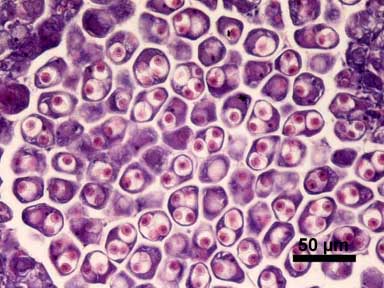
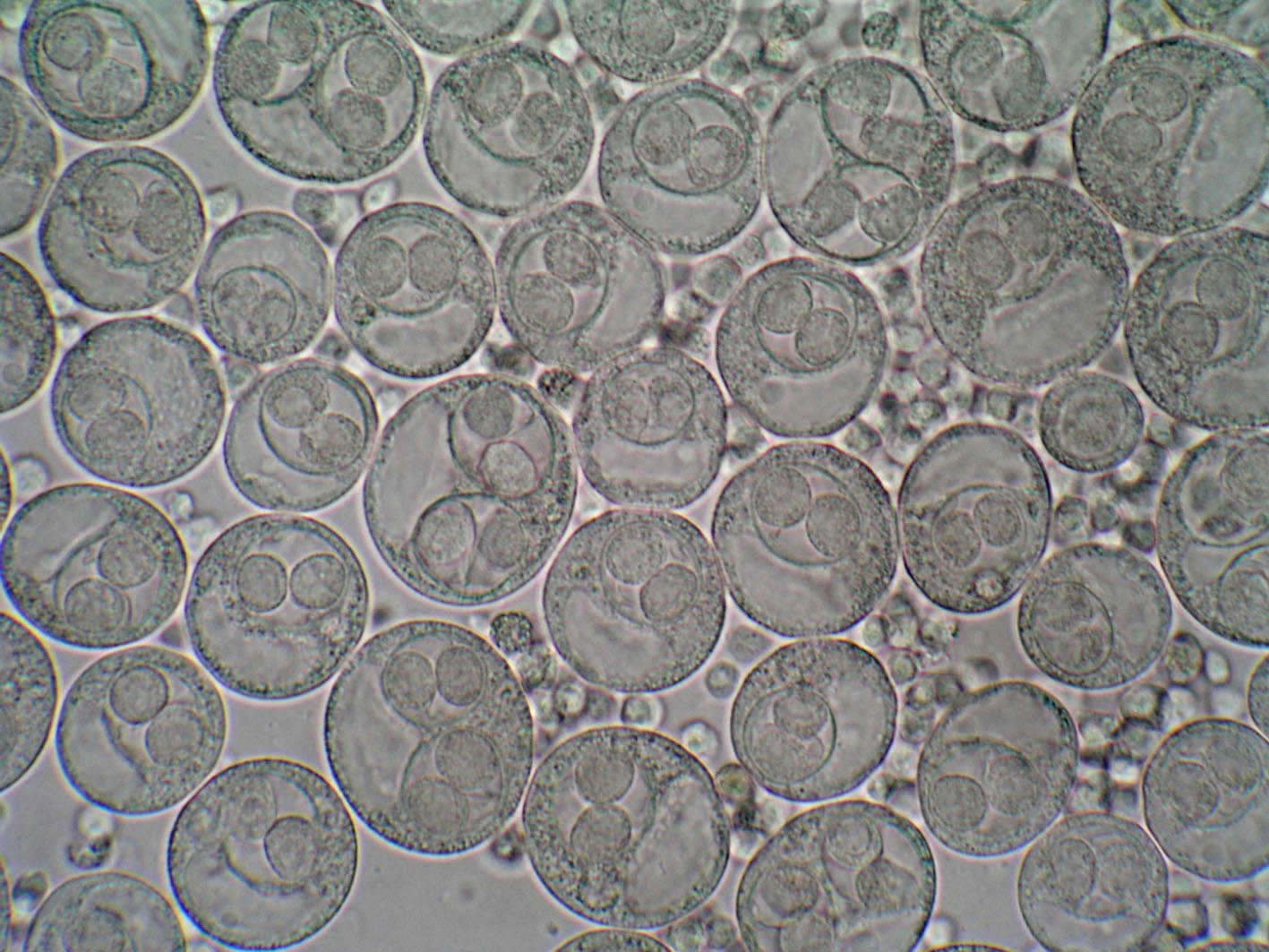
Fig. 3. Histological section of infected ovary. HE stain.
Fig. 1. Pacific oyster exhibiting the nodules in the ovary.
Fig. 2. Wet mount of infected ovary. The intracellular parasites are observed in the oocytes.