| Parasite | Myxobolus artus |
|---|---|
| Taxonomy | Myxozoa, Myxosporea, Bivalvulida |
| Host | Common carp (Cyprinus carpio) |
| Infection site | Trunk muscle |
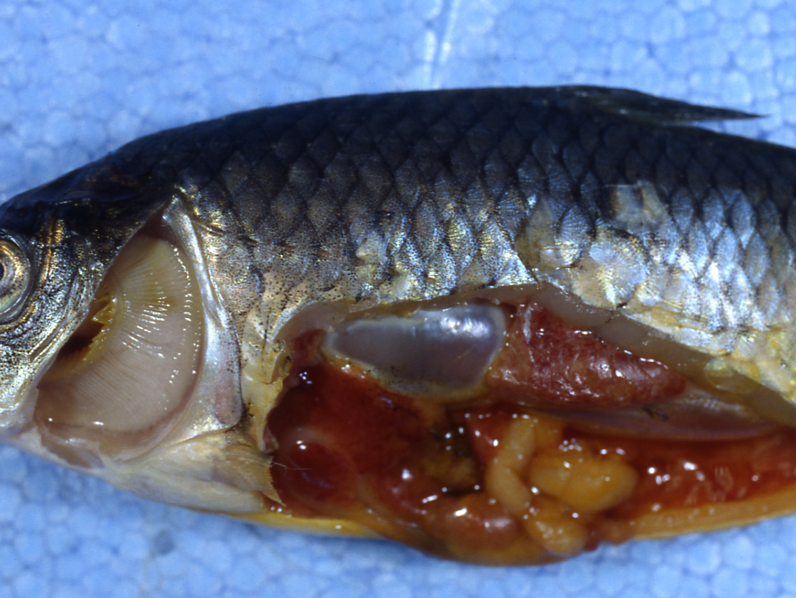
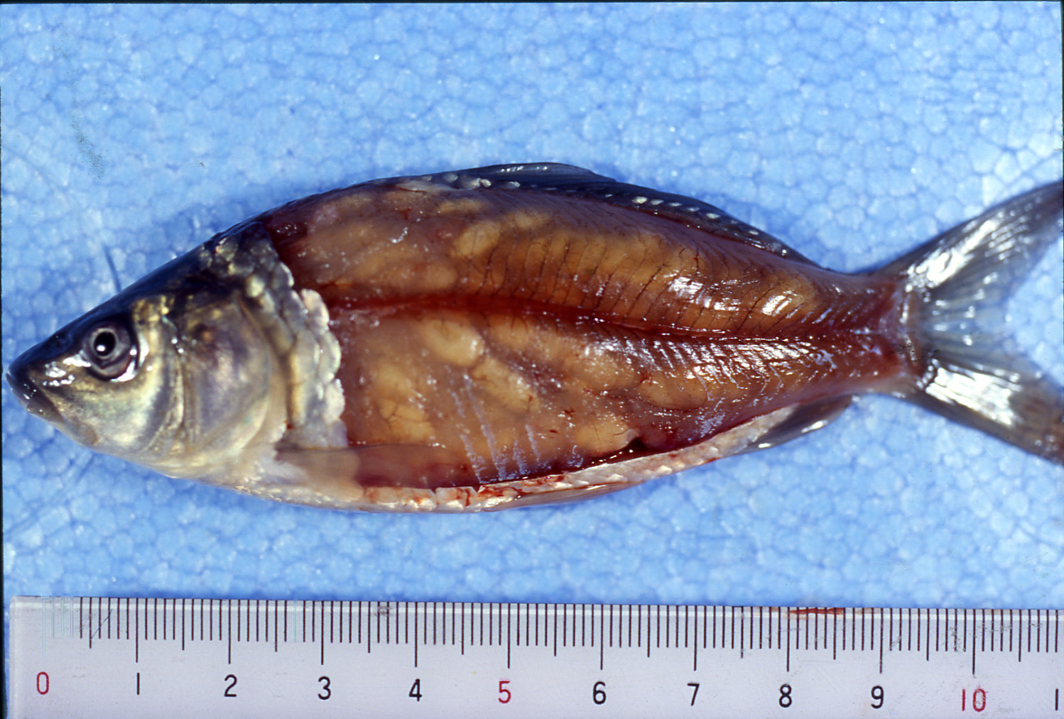
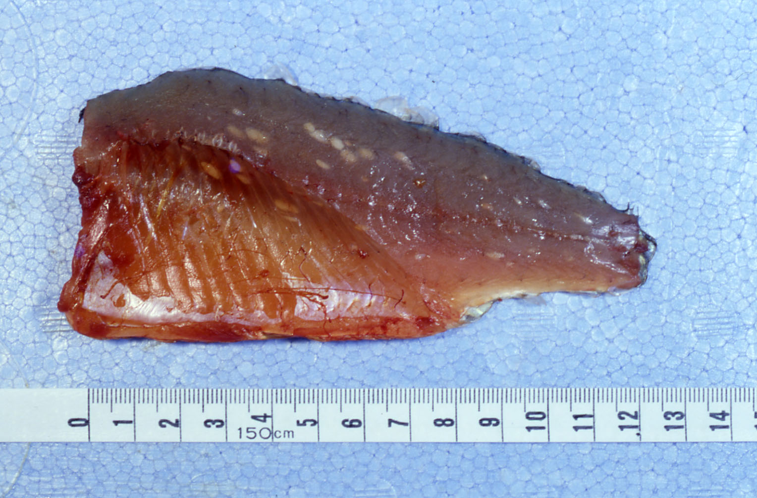
| Clinical signs | White cysts (a few mm in size) were formed in the trunk muscle. In 1-year old fish, the infection is subclinical and the macroscopic cysts in trunk muscle can be detected only after fish are filleted (Fig. 1). On the other hand, in 0-year fish, affected fish is often externally recognizable by irregular and uneven body surface (Fig. 2). In case of heavy infection, anemic gills and enlarged kidney are observed (Fig. 3). |
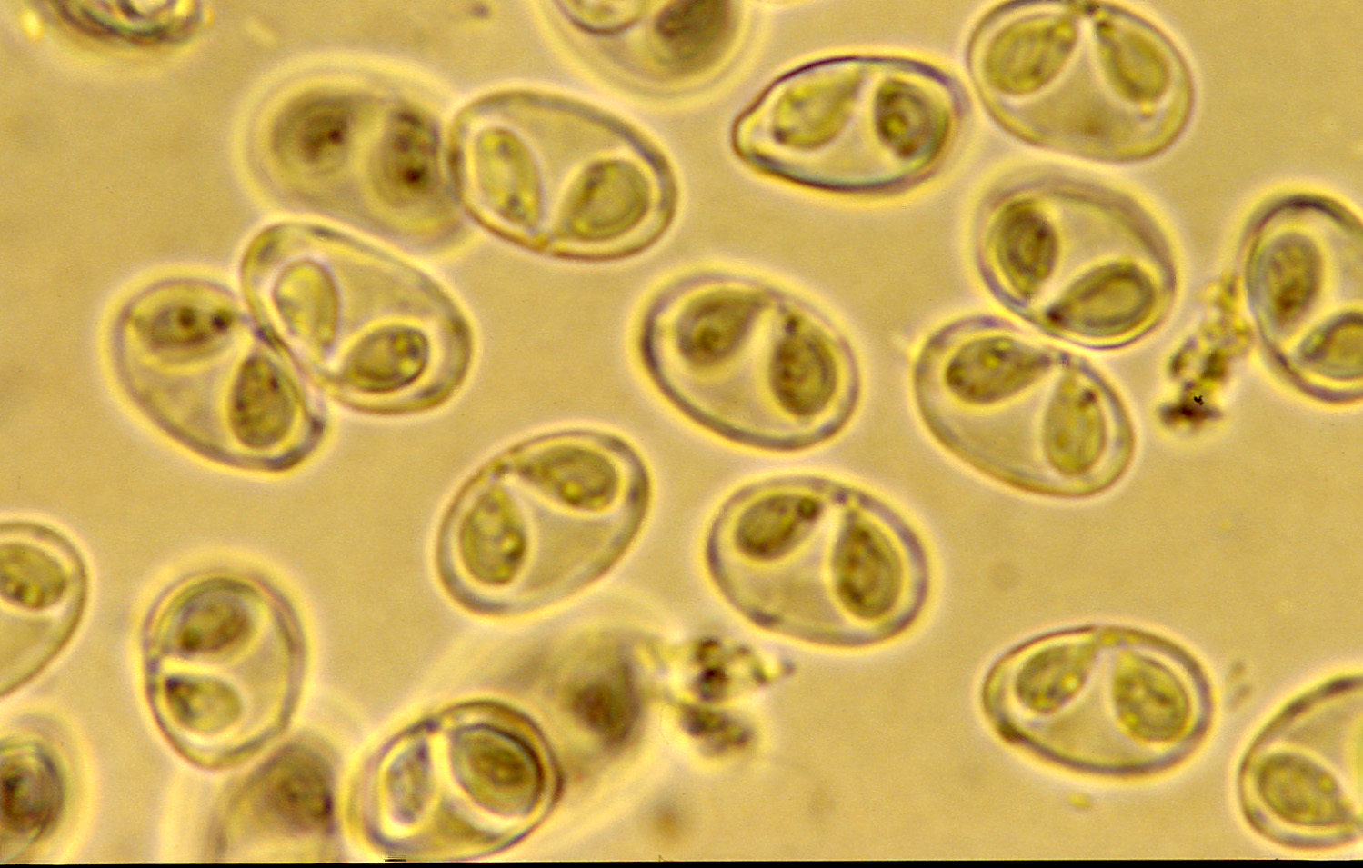
| Parasitology | Many spores are produced in cysts (Fig. 4). A spore (length 7.6-9.5 mm ;width 7.6-9.5 mm; thickness 5.7-6.3 mm) is ellipsoidal (longitudinally compressed) and has two polar capsules (length 4-5 mm; width 3-4 mm). The life cycle is unknown. The alternate host is probably involved in the life cycle. |
| Pathology | Interfibrillar cysts are encapsulated by the connective tissues of the host. In heavily infected 0-year fish, most muscle tissues are replaced by the parasite, though it doesn't result in acute mortality. After the sporulations in the cysts, the spores are dispersed following to disintegration of the atrophied connective tissues. Macrophages phagocytize spores and transportes to the kidney, spleen, etc. Accumulation in the kidney is remarkable and the enlarged kidney is sometimes visually observed. The spores transported to the intestine, the body surface and the gills are released outside the fish (Ogawa et al., 1992). Accumulation of numerous spores in the gills causes the destruction of dilated capillaries and gill epithelia followed by the hemorrhages. The diseased fish exhibites the hemorrhagic anemia characterized by the increase of the immature erythrocytes and the decreases of hematocrit value, hemoglobin contents and erythrocyte count (Yokoyama et al., 1996). |
| Health hazard | Since this parasite is not infectious to human, it is harmless in food hygiene. |
| Diagnosis | The presumptive diagnosis can be performed when cysts are observed in the muscle of the common carp. Check the spores by the wet-mount of cysts to perform the definitive diagnosis. Sample should be smeared and stained by Giemsa or Diff-Quik. |
| Other information | This disease occurred in the cultured common carp at Lake Kasumigaura and in many parts of Japan in the mid-1980s. It is considered that the fish are infected from May to June. Cysts of M. artus can be grossly observed from summer to autumn. Mature spores are released from autumn to spring. In that time, the heavily infected 0-year fish die chronically due to the hemorrhagic anemia. There are no effective methods to treat for this disease. Although the cysts in the 0-year fish gradually disappear after autumn, the fish recover over a long time. Therefore, remove the heavily infected fish immediately. It was reported that the production of antibody against the vegetative stages of Myxobolus artus when the cysts disrupt during the development, but it is unclear whether this reaction is involved in the host defense to the M. artus. (Furuta et al., 1993). |
| References | Furuta, T., K. Ogawa
and H. Wakabayashi (1993): Humoral immune response of carp Cyprinus carpio to Myxobolus
artus (Myxozoa: Myxobolidae) infection. J.
Fish Biol., 43, 441-450. Ogawa, K., K. P. Delgahapitiya, T. Furuta and H. Wakabayashi (1992): Histological studies on the host response to Myxobolus artus Akhmerov, 1960 (Myxozoa: Myxobolidae) infection in the skeletal muscle of carp, Cyprinus carpio L. J. Fish Biol., 41, 363-371. Yokoyama, H., T. Danjo, K. Ogawa, T. Arima and H. Wakabayashi (1996): Hemorrhagic anemia of carp associated with spore discharge of Myxobolus artus. Fish Pathol., 31, 19-23. |
Fig. 3. Anaemia of gill and enlarged kidney of heavily infected fish
Fig. 1. A fillet of 1-year old common carp infected with M. artus.
Fig. 2. 0-year old fish heavily infected with M. artus.

Fig. 4. Fresh spores of M. artus
(Photo by K. Ogawa (1))