| Parasite | Myxobolus episquamalis |
|---|---|
| Taxonomy | Myxozoa, Myxosporea, Bivalvulida |
| Host | Flathead mullet (Mugil cephalus) |
| Infection site | Scale |
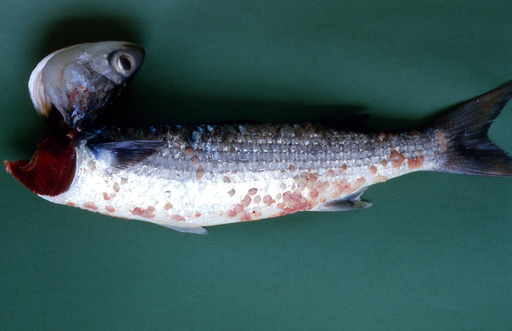
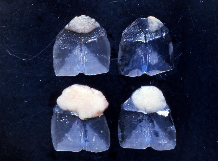
| Clinical sign | Reddish aggregates of cysts are observed in the scale (Fig. 1). The aggregates are various shaped, e.g. semicircular, rhomboid, and occupy various spaces, e.g. a part of the scale or entire surface of the scale. The infected fish exhibits ugly appearance due to the reddish foci caused by the proliferation of the blood vessels around the cysts (Fig. 2). |
| Parasitology | Many spores are produced inside the cysts (Fig. 3). A spore (length 7.5-9.5 (average 8.6) mm; width 6.0-7.5 (6.8) mm; thickness 4.5-5.5 (5.1) mm) is oval, tapering anteriorly to a blunt apex and has 2 polar capsules (length 3.8-5.0 (4.4) mm; width 2.0-3.0 (2.2) mm). The life cycle is unknown. |
| Pathology |
The aggregates of cysts were in contact with the bony plate in the scale and thesurface was covered by a thin collagenous layer, except for the marginal area which was roofed with epithelial tissues of the host. Numerous cysts existed among the proliferated connective tissue inside the aggregates. The blood vessels proliferated among the cysts caused the reddish appearance of the aggregates (Egusa et al., 1989). It is unclear whether infection is fatal or not. |
| Health hazard | Since this parasite is not infectious to human, it is harmless in food hygiene. |
| Diagnosis | Check the spores by wet-mount of cysts. Sample should be smeared and stained by Giemsa or Diff-Quik. |
| Other information | This disease has been reported from the coastal water since 1980s. Egusa et al. (1990) described this species as Myxobolus episquamalis, but some authors suggested that M. episquamalis is a synonym of Myxobolus exiguus, a parasite of mullets in the Mediterranean Sea, because of the similarities in the infection site and the morphological characteristics (Lom and Dykova, 1992). However, the molecular phylogenic analysis demonstrated that they are not the same species (Bahri et al., 2003). |
| References | Bahri, S., K. B.
Andree and R. P. Hedrick (2003): Morphological and phylogenetic studies of
marine Myxobolus spp. from mullet in Ichkeul Lake, Tunisia. J. Eukaryot. Microbiol., 50,
463-470. Egusa, S., H. Jyo, H. Oka and K. Ikata (1989): Skin disease of Mugil cephalus due to Myxobolus sp. (Myxozoa: Myxobolidae). Fish Pathol., 24, 59-60. (In Japanese) Egusa, S., Y. Maeno and M. Sorimachi (1990): A new species of Myxozoa, Myxobolus episquamalis sp. nov. infecting the scales of the mullet, Mugil cephalus L. Fish Pathol., 25, 87-91. Lom, J. and I. Dykova (1992): Protozoan parasites of fishes. Elsevier, New York, 315p. |

Fig. 2. Scales of infected mullet. White to pink regions are aggregates of the parasite.
Fig. 3. Fresh spores of M. episquamalis
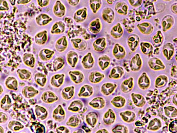
(Photos by K. Momoyama)
Fig. 1. Diseased flathead mullet