
| Parasite | Microsporidium sp. |
|---|---|
| Taxonomy | Microspora, Microsporea |
| Hosts | Penaeid shrimps (Trachypenaeus curvirostris, Metapenaeopsis barbata, Metapenaeus ensis) |
| Infection site | Muscle |
| Clinical sign | The muscle lesion is whitish like heat-processed muscle (Fig. 1). Infected Trachypenaeus curvirostris exhibits a loss of shiny body surface, reddish body and darken dorsal region. |
| Parasitology | A number of spores fill up the muscle tissue (Fig. 2). A spore is ovoid and 1.8-2.0 mm long and 1.0-1.2 mm wide. |
| Pathology | No report |
| Health hazard | Since this parasite is not infectious to human, it is harmless in food hygiene. |
| Diagnosis | Check the spores by wet-mount of cysts. The sample should be smeared and stained by Uvitex 2B followed by a fluorescent microscopic observation. The stained spores emit blue fluorescence under UV radiation. |
| Other information | The activity of infected prawn was almost similar to non-infected prawn, while the former shows a poor durability (Momoyama and Tensha, 2006). |
| References | Momoyama, K. and K. Tensha (2006): Ugly-looking parasitic infections and other abnormalities of wild fish and shellfish caught in the coastal or inland waters around or in Yamaguchi Prefecture. Bull. Yamaguchi Pref. Fish. Res. Ctr., 4, 143-161.(In Japanese) |
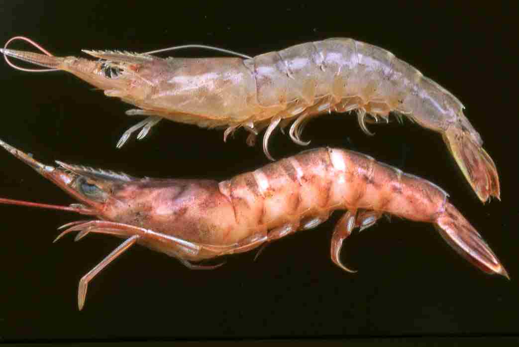
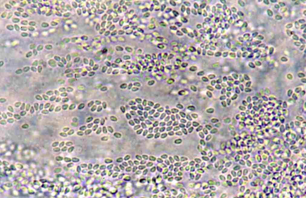
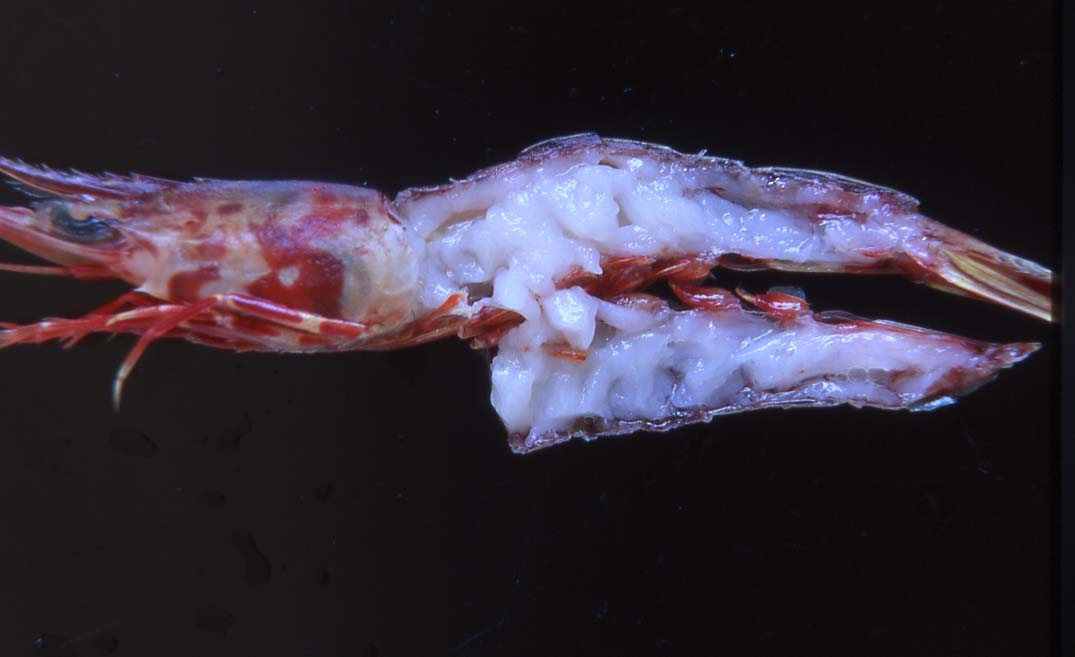

Fig. 3. Discoloured muscle tissue of infected prawn.
Fig. 1. External appearance of healthy (above) and diseased (below)
shrimps.
(Photos by K. Momoyama)
Fig. 2. Muscle of infected (above) and non-infected (below) shrimp.
Fig. 4. Fresh spores of Microsporidium sp. from the muscle tissue.