| Parasite | Microsporidium sp. |
|---|---|
| Taxonomy | Microspora, Microsporea |
| Host | Kuruma prawn (Penaeus japonicus) |
| Infection site | Connective tissue under the exoskeleton, nervous tissue. |
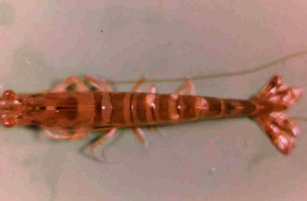
| Clinical sign | The lesion appears swollen since white aggregates of microsporidial cysts are formed in the connective tissue under the exoskeleton (Fig. 1). |
| Parasitology | Many spores are formed inside the cysts (Fig. 2). A spore (length ca. 3.9 mm; width ca. 2.0 mm) is ovoid. This microsporidian is possibly a new species because the infection site is different from that of the microsporidian reported in the kuruma prawn previously. |
| Pathology | The rate of mortality was higher in infected larvae than non-infected larvae (Momoyama and Tensha, 2006). |
| Health hazard | Since this parasite is not infectious to human, it is harmless in food hygiene. |
| Diagnosis | Check the spores by wet-mount of cysts. The sample should be smeared and stained by Uvitex 2B followed by a fluorescent microscopic observation. The stained spores emit blue fluorescence under UV radiation. |
| Other information | This parasite may be pathogenic because of parasitizing the nervous tissue. However, the escape response of the infected prawn was not so impaired (Momoyama and Tensha, 2006). |
| References | Momoyama, K. and K. Tensha (2006): Ugly-looking parasitic infections and other abnormalities of wild fish and shellfish caught in the coastal or inland waters around or in Yamaguchi Prefecture. Bull. Yamaguchi Pref. Fish. Res. Ctr., 4, 143-161 (In Japanese). |
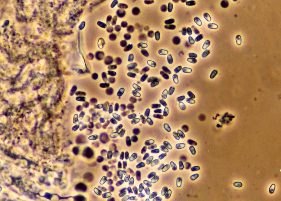

Fig. 1. White cysts are observed under the dorsal exoskeleton of
kuruma prawn.
(Photos by K. Momoyama)
Fig. 2. Fresh spores of Micrsporidium sp.