| Parasite | Microsporidium sp. |
|---|---|
| Taxonomy | Microspora, Microsporea |
| Host | Red sea bream (Pagrus major) |
| Disease name | Beko disease ('Beko' means uneven , concave surface in Japanese.) |
| Infection site | Trunk muscle |
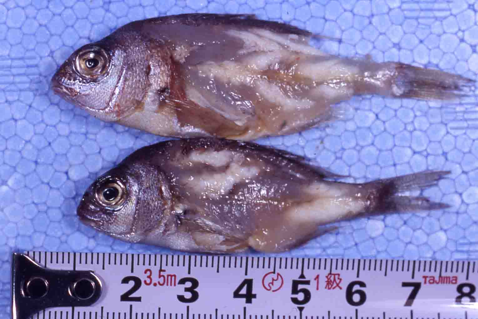
| Clinical signs | Infected fish exhibits uneven and concave body surface. White cysts are observedin trunk muscle (Fig. 1). |
| Parasitology | Many spores are produced inside the cysts. The spore of this parasite (length 2.9-3.9mm; width 1.9-2.6 mm) is slightly larger than that of Microsporidium seriolae, infecting to yellowtail (Egusa et al., 1988). |
| Pathology | Histopathological characteristics and the development of the parasite are similar to those in beko disease in yellowtail. Cysts are encapsulated by the host connective tissues and the parasite proliferates and sporulates inside the cysts. |
| Health hazard | Since this parasite is not infectious to human, it is harmless in food hygiene. |
| Diagnosis | Check the spores by the wet-mount of cysts. The sample should be smeared and stained by Uvitex 2B followed by a fluorescent microscopic observation. The stained spores emit blue fluorescence under UV radiation. |
| Other information | This disease occurs only in juvenile stage of red sea bream. Usually, fish recovers after release of spores outside the host. However, cysts in trunk muscle sometimes remain even in commercial-sized fish. |
| References | Egusa, S., K. Hatai
and Y. Fujimaki (1988): Notes on Microsporidium
species, the
etiological agent of “Beko” disease in red sea bream juveniles, Pagrus major. Fish Pathol., 23, 263-267. (In Japanese) |

Fig. 1. Microsporidium cysts in the muscle of red sea bream fry.