| Parasite | Myxobolus koi |
|---|---|
| Taxonomy | Myxozoa, Myxosporea, Bivalvulida |
| Host | Common carp (Cyprinus carpio) |
| Infection site | Gill |
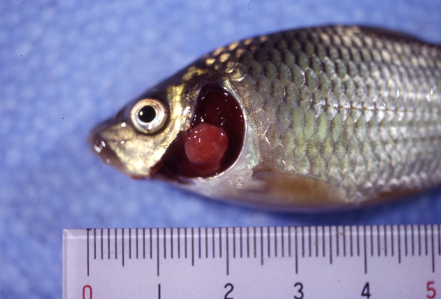
| Clinical signs | White cysts (a few mm in size) are observed in the gill of common carp fry. Diseased fish exhibits the secretion of mucus, the hemorrhage in the gill, and the loss of a part of the gill filament (Fig. 1). Heavily infected fish are lethargic and piping for air. Two types of cysts are often observed; one is a large-type and the other is a small-type cyst with a diameter of below 1 mm (Fig. 2). |
| Parasitology | Many spores are produced inside the cysts (Fig. 3). A spore (length 12-15 mm; width 5-9 mm; thickness 5-8 mm) is oval to pyriform and has 2 equal polar capsules (length 5-8 mm; width 1.6-2.7 mm). The life cycle is unknown. Alternate host is probably involved in the life cycle. |
| Pathology | The parasite develops inside the gill filament and forms the complex cysts, encasing the gill tissues. As a result, the gills are congested by the occlusion of the blood capillaries and the remarkable host reactions such as the proliferations of gill epithelia or the fusion of gill filaments (Fig. 4). Oxygen deficiency in the early summer causes a mass mortality of heavily infected fish (Egusa, 1988). It is considered that the formations of large-type cysts are fatal to host fish, while small-type cysts are not harmful. |
| Health hazard | Since this parasite is not infectious to human, it is harmless in food hygiene. |
| Diagnosis | Check the spores by wet-mount of the cysts. M. koican be distinguished from Myxobolus toyamai and Myxobolus musseliusaeby spore morphology. M. toyamai is slender pyriform and has unequal 2 polar capsules; M. musseliusae is spherical or ovoid. Sample should be smeared and stained by Giemsa or Diff-Quik. |
| Other information | This disease has been frequenly found in the culture of common carp and called as “cheek swelling” since the opercle is swollen up. Egusa (1988) suggested that spores from large-type cysts and those from small-type cysts are different species because of the slight differences in spore shape and spore size. However, Yokoyama et al. (1997) elucidated by the detailed morphological analysis and the indirect fluorescent antibody technique that these spores derived from the same species. Although there are no effective control methods, diseased fish recover after spore release in autumn. Therefore, it is important to prevent mortality by oxygen deficiency in summer (Yokoyama, 2004). |
| References | Egusa, S. (1988): Protozoan
diseases. Fish pathology (Egusa, S.), Koseisha
koseikaku, pp. 219-274. Yokoyama, H. (2004): Myxozoan diseases. Infectious and parasitic diseases of fish and shellfish. (Wakabayashi, H. and K. Muroga), Koseisha koseikaku, pp.339-352. Yokoyama, H., D. Inoue, A. Kumamaru and H. Wakabayashi (1997): Myxobolus koi (Myxozoa: Myxosporea) forms large-and small-type ‘cysts’ in the gills of common carp. Fish Pathol., 32, 211-217. |
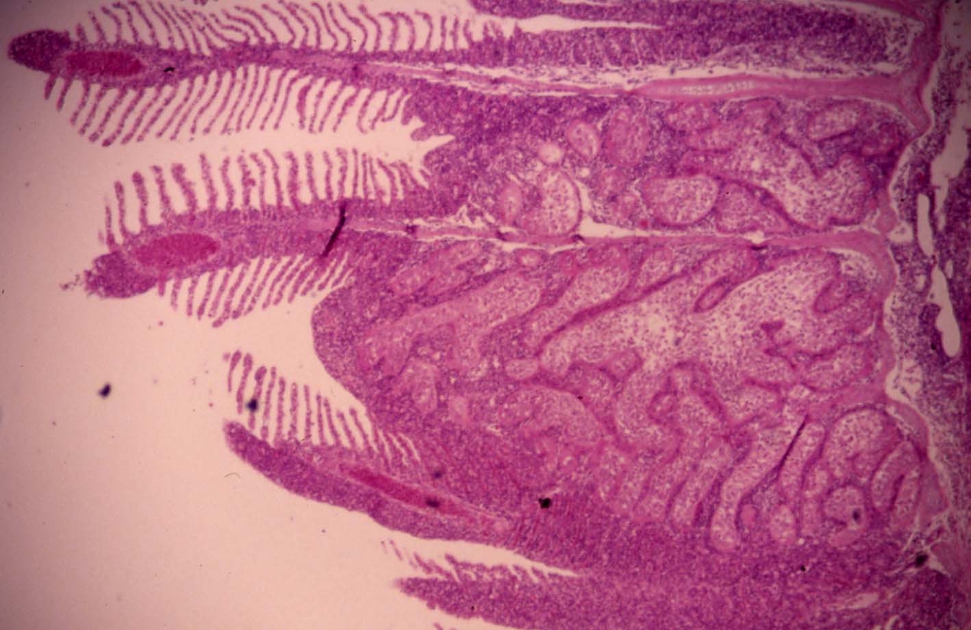
Fig. 3. Fresh spores ofM. koi
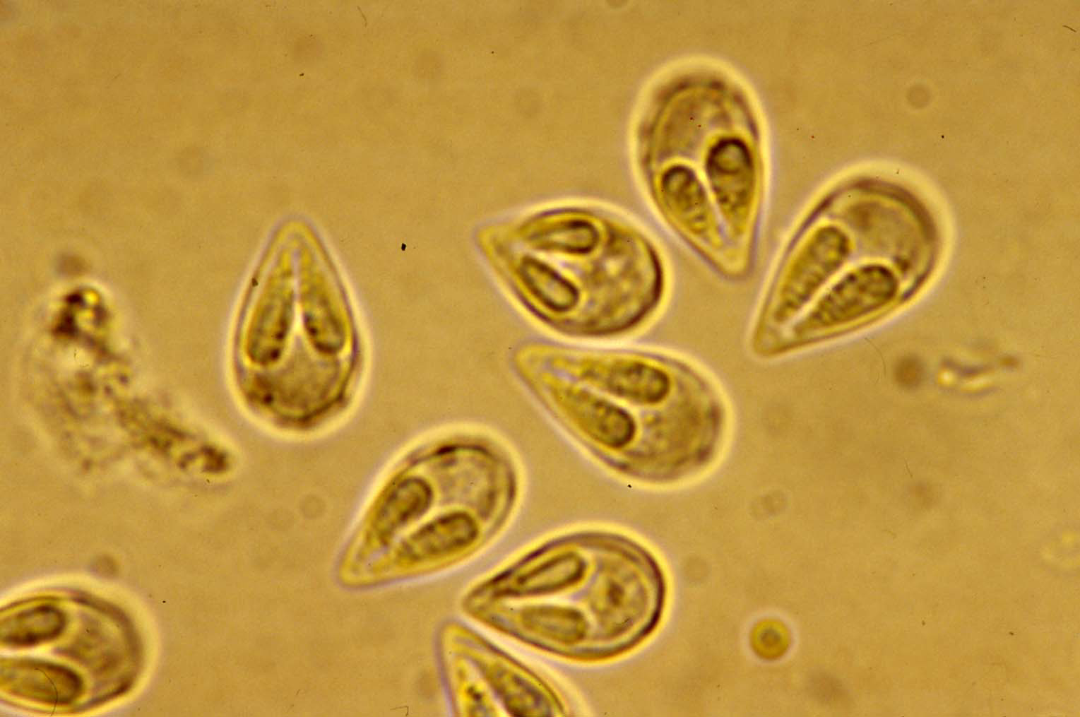
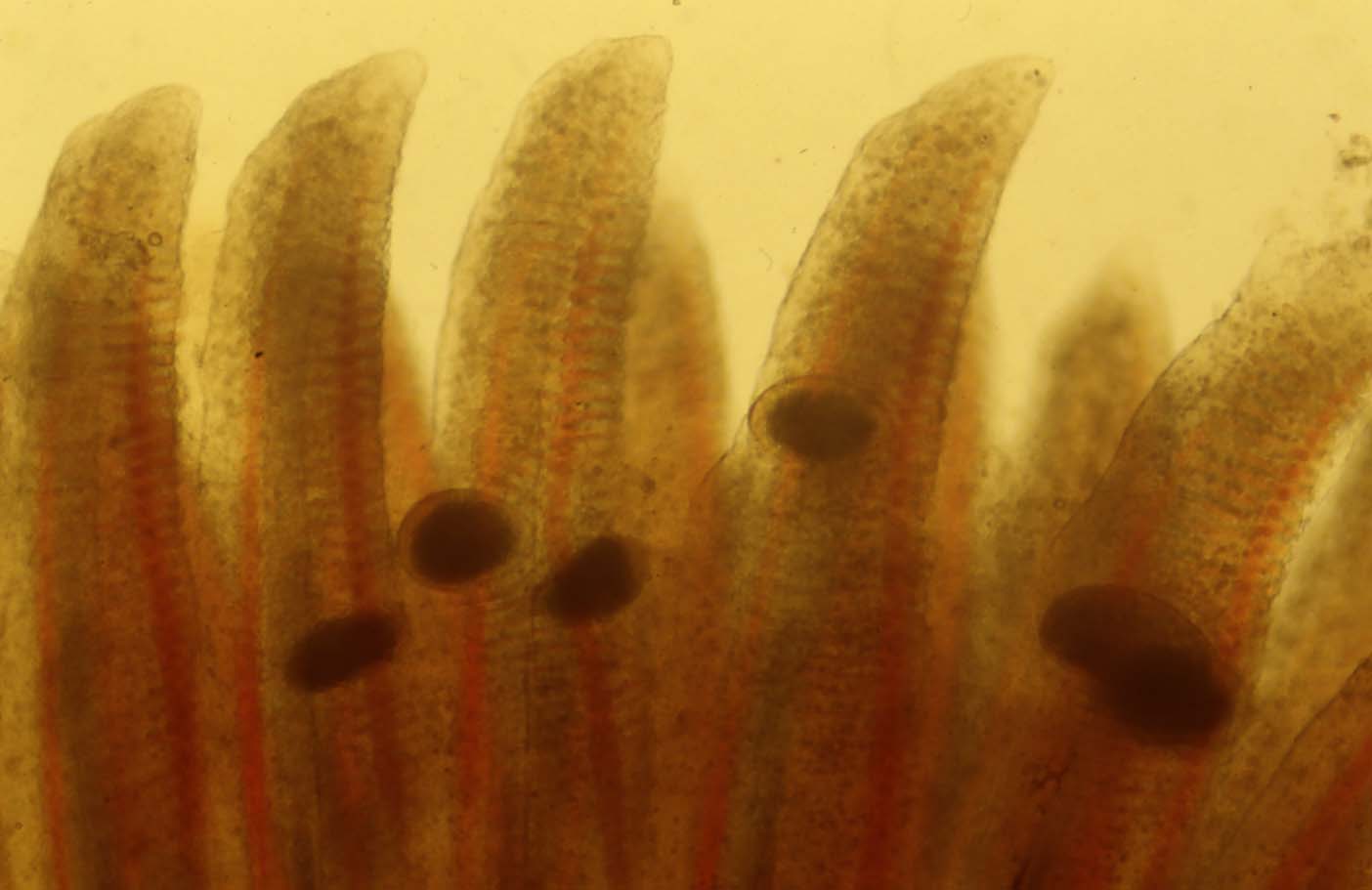

Fig. 2. Small-type cysts of M. koi in the gill filament.
Fig. 1. A large-type cyst of M. koi in common carp fry.
Fig. 4. Histology of Large-type cyst of M. koi.