| Parasite | Myxobolus nagaraensis |
|---|---|
| Taxonomy | Myxozoa, Myxosporea, Bivalvulida |
| Hosts | Rhinogobius sp. OR, Rhinogobius flumineus, Rhinogobius sp. CB |
| Infection site | Body cavity, caudal peduncle |
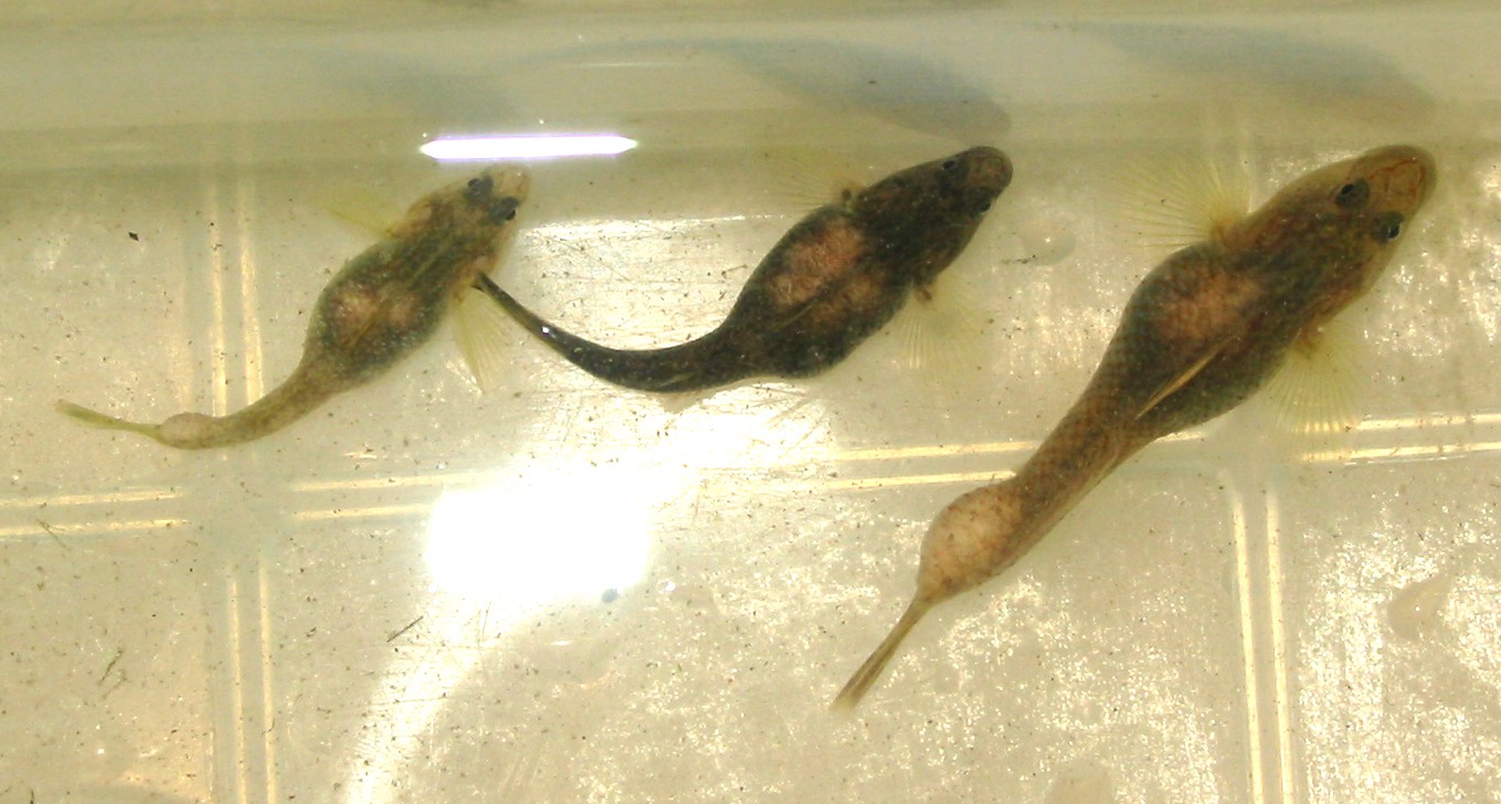
| Clinical signs | Diseased fish exhibits the abdominal distension and the nodules of the caudal peduncle (Fig. 1). Large parasitic cysts, aggregates of several smaller cysts, are observed (Fig. 2). |
| Parasitology | Many spores are produced inside the cysts (Fig. 3). A spore (length 10.5-13.5 (average 11.9) mm; width 8.0-10.0 (9.0) mm; thickness 6.0-7.0 (6.5) mm) is ovoid and has 2 polar capsules (length 4.5-6.0 (5.5) mm; width 2.5-4.0 (3.0) mm). The life cycle is unknown. |
| Pathology |
In the early stage of infection, the parasite develops between the connective tissue of the renal capsule. Aggregates of plasmodia are encased by the host's connective tissues, resulting in formation of cysts. The parasitic masses extend into the visceral cavity and force the internal organs (Yokoyama et al., 2007). Occasionally, the cysts also occur in the caudal peduncle. It is unclear whether this disease could kill the host fish. |
| Health hazard | Since this parasite is not infectious to human, it is harmless in food hygiene. |
| Diagnosis | Check the spores by wet-mount of cysts. Sample should be smeared and stained by Giemsa or Diff-Quik. |
| Other information | Myxobolus nagaraensis was found in Rhinogobius spp. collected from a tributary of the Nagara River in Gifu prefecture. |
| References | Yokoyama, H., T. Kageyama, K. Ohara and T. Yanagida (2007): Myxobolus nagaraensis n. sp. (Myxozoa: Myxospora) causes abdominal distension of freshwater goby Rhinogobius sp. OR type from the Nagara River. Fish. Sci., 73, 633-639. |
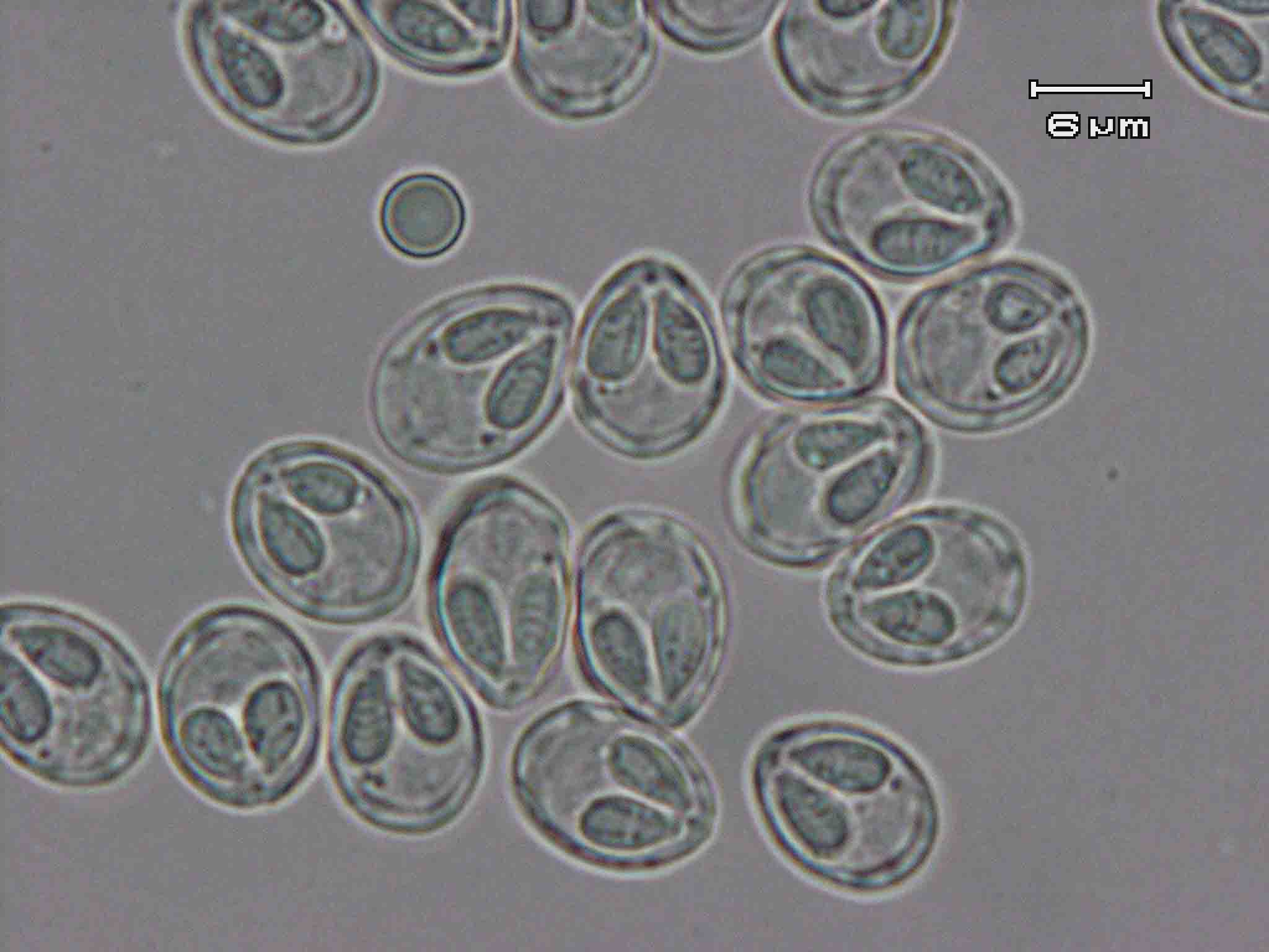
Fig. 2. Cysts of M. nagaraensis in the body cavity and the caudal
peduncle.
Fig. 1. Dorsal view of infected Rhinogobius.
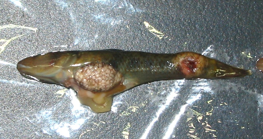

Fig. 3. Fresh spores of M. nagaraensis