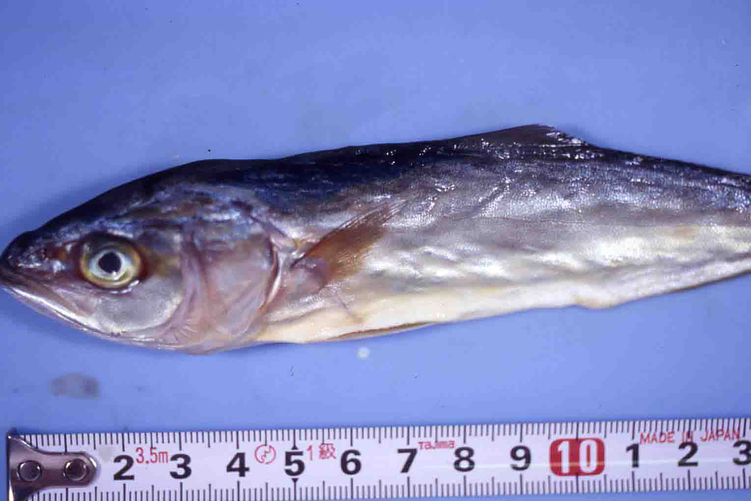
Fig. 1. External appearrance of diseased yellowtail fry.
Fig. 5. M. seriolae spores stained with Uvitex 2B.
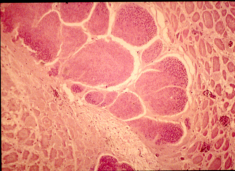
Fig. 4. Histology of cyst of M. seriolae in the trunk muscle
Fig. 2. Mass of Microsporidium cysts in the trunk muscle.
| Parasite | Microsporidium seriolae |
|---|---|
| Taxonomy | Microspora, Microsporea |
| Hosts | Yellowtail (Seriola quinqueradiata), Greater amberjack (Seriola dumerili), Yellowtail amberjack (Seriola lalandi) |
| Disease name | Beko disease ('Beko' means an uneven and concave surface in Japanese.) |
| Infection site | Trunk muscle |
| Clinical signs | Infected fish have small cyst-like bodies (a few mm-1 cm) in the muscle and show the concave body surface (Fig. 1). The white ‘cysts’ exhibit the variable shape (Fig. 2). |
| Parasitology | Many spores are formed in the cyst (Fig. 3). Spore length 2.9-3.7 mm; spore width 1.9-2.4 mm. The life cycle is unknown. This parasite doesn't transmit from fish to fish, suggesting that an intermediate host is involved in the life cycle. |
| Pathology | The cyst, in which the parasites proliferate and sporulate, is encapsulated by the thin connective tissue of host (Fig. 5). Few host reactions are observed during the cyst development. After cyst's degeneration, the neighboring muscle tissue shows diffuse colliquative necrosis, causing the concave body surface. Heavy infection often leads fish to death (Ogawa, 2004). After spores are ingested by phagocytes and shed into outside of the host, the fish may be recovered (Sano et al., 1998). |
| Health hazard | Since this parasite is not infectious to human, it is harmless in food hygiene. |
| Diagnosis | Check the spores by wet-mount of cysts. The sample should be smeared and stained by Uvitex 2B followed by a fluorescent microscopic observation (Yokoyama et al., 1996). The stained spores emit blue fluorescence under UV radiation (Fig. 4). The high sensitive detection method by PCR was developed (Bell et al., 1999), although its specificity is uncertain. |
| Other information | This disease occurs in juvenile fish just after they are reared in net cage. In a land-based tank, sand filtration of sea water is effective for removing the infective agent (Sano et al., 1998). |
| References | Bell.
A. S., H. Yokoyama, T. Aoki, M. Takahashi and K. Maruyama (1999): Single and
nested polymerase chain reaction assays for the detection of Microsporidium seriolae (Microspora),
the causative agent of ‘Beko’ disease in yellowtail Seriola quinqueradiata. Dis.
Aquat. Org., 37, 127-134. Ogawa, K. (2004): Protozoan diseases. Infectious and parasitic diseases of fish and shellfish. (ed. by Wakabayashi, H. and K. Muroga), Koseisha koseikaku, pp.285-338. (In Japanese) Sano, M., J. Sato and H. Yokoyama (1998): Occurrence of beko disease caused by Microsporidium seriolae (Microspora) in hatchery-reared juvenile yellowtail. Fish Pathol., 33, 11-16. Yokoyama, H., J.-H. Kim, J. Sato, M. Sano and K. Hirano (1996): Fluorochrome Uvitex 2B stain for detection of the microsporidian causing beko disease of yellowtail and goldstriped amberjack juveniles. Fish Pathol., 31, 99-104. |

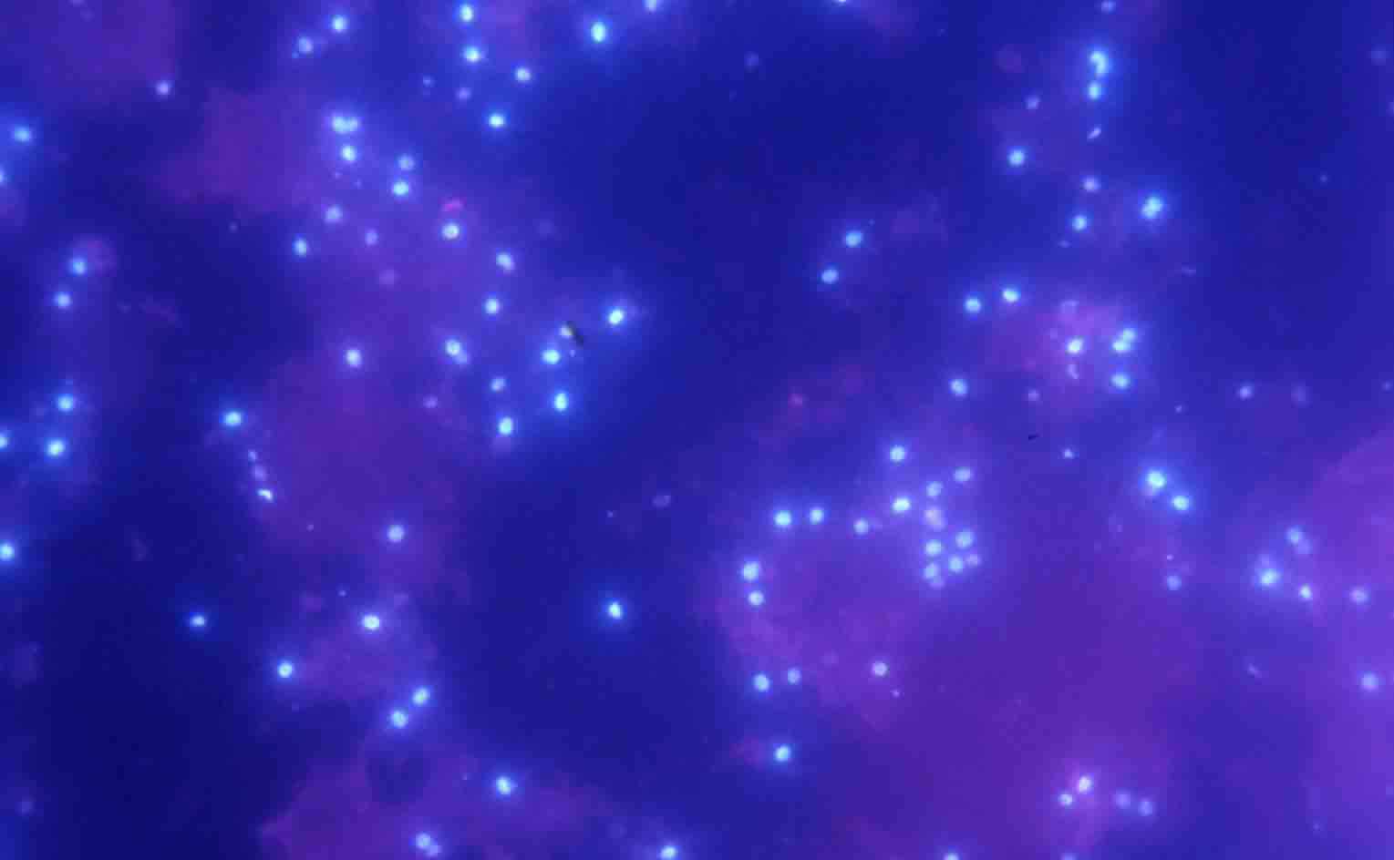
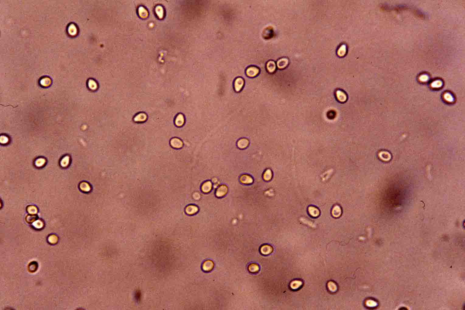
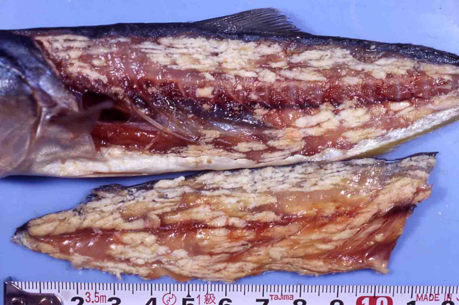
Fig. 3. Fresh spores of Microsporidium seriolae