| Parasite | Kudoa sp. |
|---|---|
| Taxonomy | Myxozoa, Myxosporea, Multivalvulida |
| Host | North-Pacific giant octopus (Paroctopus dofleini) |
| Infection site | Muscle in the arm. |
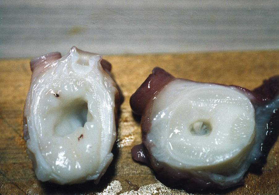
| Clinical sign | The central region of the arm is liquefied, resulting in a large hole by the muscle breakdown (Fig. 1). |
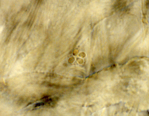
| Parasitology | A spore is observed in the liquefied muscle tissue (Fig. 2). A spore (6-8 mm in diameter) has 4 polar capsules (ca. 2.5×1.5 mm). Life cycle is unknown. |
| Pathology | A post-mortem myoliquefaction may be caused by the proteolytic enzymes derived from the parasite. |
| Health hazard | Since this parasite is not infectious to human, it is harmless in food hygiene. |
| Diagnosis | Check the spores by wet-mount of the liquefied muscle. Sample should be smeared and stained by Giemsa or Diff-Quik. |
| Other information | An abnormal texture in the basal arm of a boiled north-Pacific giant octopus was found in a octopus processing plant. This parasite is only the species reported in octopus (Yokoyama and Masuda, 2001). |
| References |
Yokoyama, H. and K. Masuda (2001): Kudoa sp. (Myxozoa) causing a post-mortem myoliquefaction of North-Pacific giant octopus Paroctopus dofleini (Cephalopoda: Octopodidae). Bull. Eur. Ass. Fish Pathol., 21, 266-268. |

(Photos by K. Masuda)
Fig. 1. Liquefied (left) and healthy (right) arms of octopus.
Fig. 2. A Kudoa spore detected in the liquefied lesion.