| Parasite | Paradeontacylix grandispinus, Paradeontacylix kampachi |
|---|---|
| Taxonomy | Platyhelminthes, Trematoda, Strigeidida |
| Host | Greater amberjack (Seriola dumerili) |
| Disease name | Blood fluke disease |
| Infection site | Blood vessels from heart to gill |
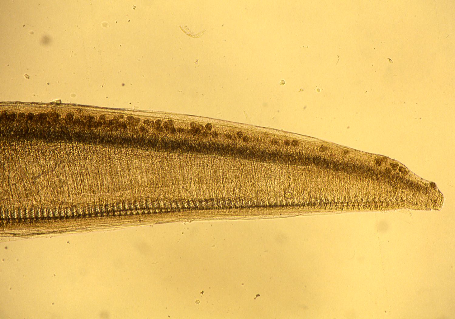
| Clinical signs | Infected fish usually exhibits no external abnormality. Heavily infected fish sometimes dies from suffocation in blockage of the gill blood vessels by the parasite eggs. Such dead fish opens its mouth. It is difficult to detect the parasite macroscopically. Parasite eggs are observed in the gill by microscopy (Fig. 1). |
| Parasitology | Both Paradeontacylix grandispinus and P. kampachi are blood flukes of Greater amberjack. The adult of the former mostly infected the afferent branchial arteries, rarely the sinus venosus and heart ventricle, while the latter is observed in various sites, including sinus venosus, heart ventricle, heart atrium, ventral aorta and afferent branchial arteries (Ogawa et al., 1993). The body (P. grandispinus: length 2.0-3.3 mm; P. kampachi: 4.7-8.1 mm) is flat and elongated (like a leaf of a willow) (Fig. 2). The intermediate host is probably involved in the life cycle, but details are unknown. |
| Pathology | Infected fish exhibits circulatory impairment due to occlusion of the branchial vessels with parasite eggs. Heavily infected fish sometimes dies from suffocation since it moves around rapidly when feeding. Eggs accumulated in the gill are encapsulated by host’s tissues to form nodules. Papillate projections are observed in the afferent branchial artery walls due to proliferations of endothelial cells (Ogawa et al., 1989). |
| Health hazard | Since this parasite is not infectious to human, it is harmless in food hygiene. |
| Diagnosis | Parasite eggs in the blood vessels can be readily found in the gills by microscopy. However, the eggs cannot be utilized for the differentiation of 2 blood flukes. Therefore, observation of adult parasites is needed for the definitive diagnosis. P. grandispinus is small (ca. 2-3 mm) compared to P. kampachi (ca. 5-8 mm) (Fig. 3). |
| Other information | Generally, heavy infection occurs in 0-yeared fish. According to Ogawa et al. (1993), the parasites invaded fish in September, the egg accumulation started in November and mortalities were suppressed in next May. On the other hand, the Greater amberjack transported from China died in May to June (Ogawa and Fukudome, 1994). Although no control methods have been reported, decreased feeding is possibly effective for this disease because heavily infected fish dies from suffocation caused by the rapid moving. |
| References | Ogawa, K. and M.
Fukudome (1994): Mass mortality caused by blood fluke (Paradeontacylix) among amberjack (Seriola dumerili) imported to Japan. Fish Pathol., 29,
265-269.
Ogawa, K., K. Hattori, K. Hatai and S. Kubota (1989): Histopathology of cultured marine fish, Seriola purpurascens (Carangidae) infected with Paradeontacylix spp. (Trematoda: Sanguinicolidae) in its vascular system. Fish Pathol., 24, 75-81. Ogawa, K., H. Andoh and M. Yamaguchi (1993): Some biological aspects of Paradeontacylix (Trematoda: Sanguinicolidae) infection in cultured marine fish Seriola dumerili. Fish Pathol., 28, 177-180. |
(Photos by K. Ogawa)

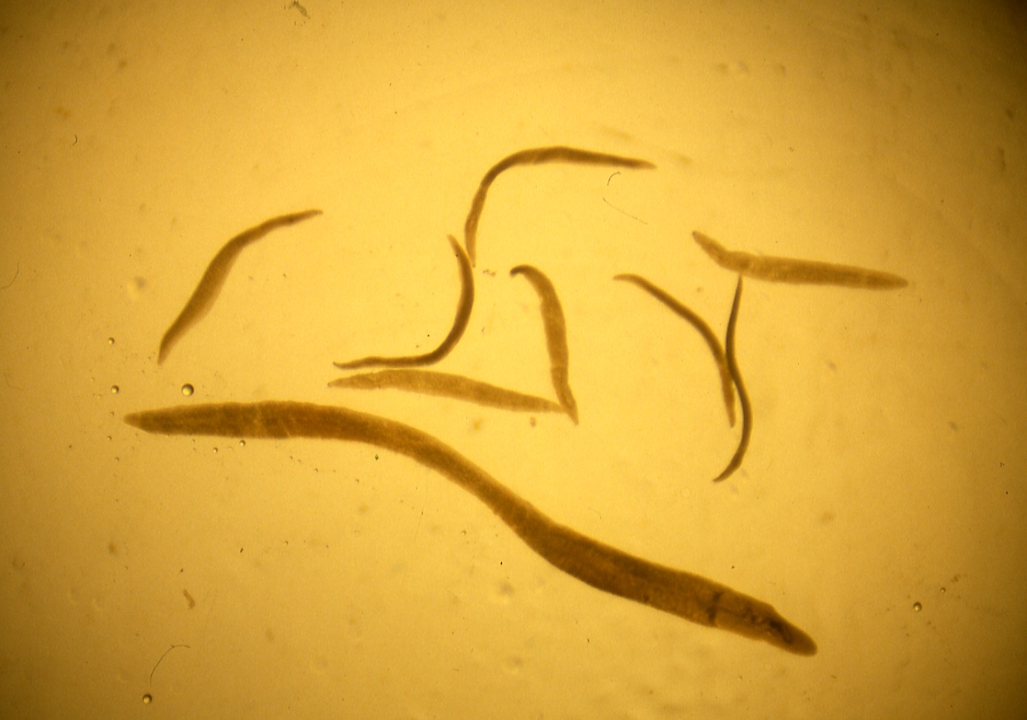
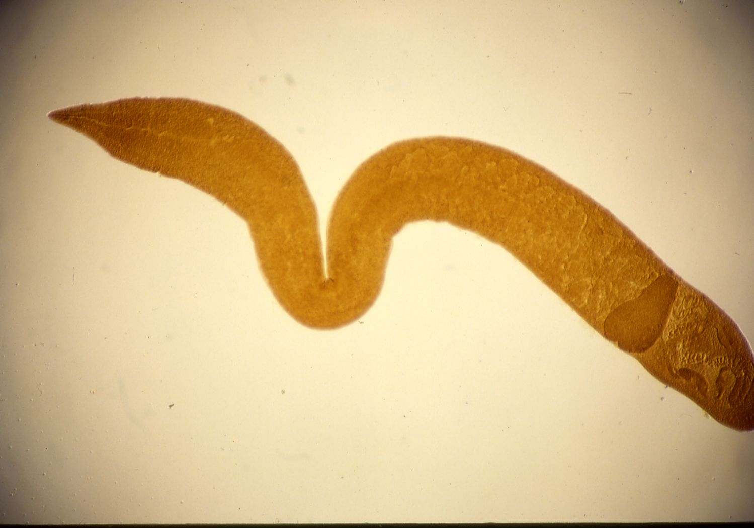
Fig. 2. Paradeontacylix kampachi
Fig. 1. Numerous eggs of blood flukes blocked in the gill capillary
of Greater amberjack..
Fig. 3. A big worm is P. kampachi and small worms are P. grandispinus