| Parasite | Rhadinorhynchus selkirki |
|---|---|
| Taxonomy | Acanthocephala, Palaeacanthocephala, Echinorhynchida |
| Host | Pacific saury (Cololabis saira) |
| Infection site | Intestine |
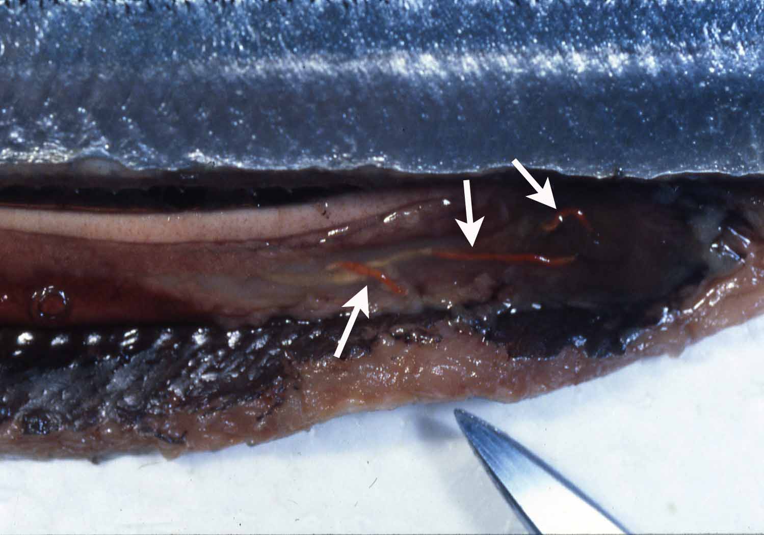
| Clinical sign | Infected fish usually exhibits no external abnormality, but rarely a part of the parasite can be seen from the anus of host fish. An elongate orange parasite is observed in the intestine (Fig. 1). |
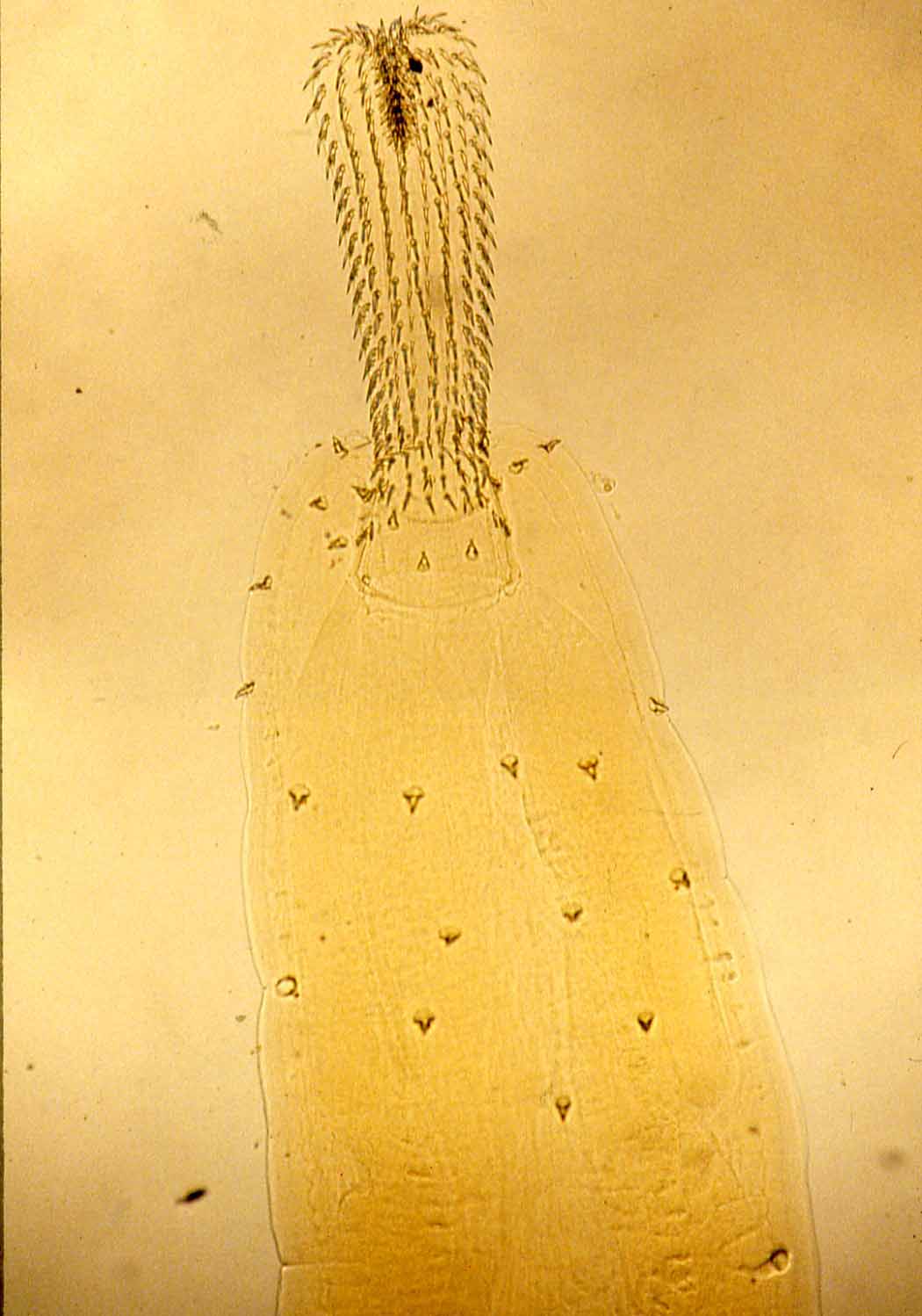
| Parasitology | The body (length 2-3 cm) is cylindrical and composed of proboscis, neck and trunk (Fig. 2). The parasite burrows into the rectum wall of the host with its proboscis and neck, and uptakes nutrients through its body surface. Dioecious. Larvae are hatched from eggs in the intermediate host (crustacean) and grow to be encysted cystacanth. |
| Pathology | Pathogenicity to host fish is low. |
| Health hazard | Since this parasite is not infectious to human, it is harmless in food hygiene. |
| Diagnosis | Orange colored appearance of the parasite body can be used for the presumptive diagnosis. Check the morphology of the proboscis for the definitive diagnosis. |
| Other information | Though this parasite occurs commonly in Pacific saury, complaints have been frequently made by consumers, due to Japanese habit of eating the viscera. R. selkirki is found in Pacific saury, while R. katsuwonis is often observed in skipjack tuna and chub mackerel. |
(Photos by K. Ogawa)
Fig. 2. The proboscis with many spines of Rhadinorhynchus.

Fig. 1. Rhadinorhynchus (arrows) in Pacific saury.