| Parasite | Spraguea americana |
|---|---|
| Taxonomy | Microspora, Microsporea |
| Host | Japanese anglerfish (Lophius litulon) |
| Infection site | Nervous tissue |
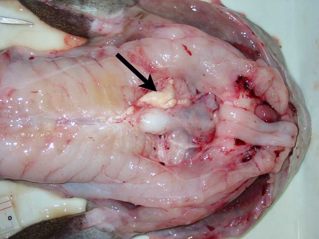
| Clinical sign | No clinical signs are evident. Groups of the white cysts are observed in various parts of nerve tissues (Figs. 1, 2). |
| Parasitology | Many elongate oval spores (3.4 x 1.8 mm) are produced inside xenomas (Fig. 3). No dimorphism of spores is observed. |
| Pathology | Spraguea americana, an intracellular parasite, infects to the host ganglion cells, forming a hypertrophied xenoma, in which many spores are produced. |
| Health hazard | Since this parasite is not infectious to human, it is harmless in food hygiene. |
| Diagnosis | Check the spores by wet-mount of cysts. Sample should be smeared and stained by Uvitex 2B followed by a fluorescent microscopic observation. The stained spores emit blue fluorescence under UV radiation. |
| Other information | This parasite was previously identified as Glugea americanus, a parasite infecting to the nervous tissues of American angler (Lophius americanus). Subsequently, molecular analysis suggested that G. americanus should be transferred to the genus Spraguea and be reassigned as Spraguea americana. S. americana is considered as a separate species from S. lophii, a parasite infecting to angler Lophius piscatorius, due to the morphological difference in spore dimorphism of the latter species, even though their gene sequences are almost identical (Freeman et al., 2004). |
| References | Freeman,M. A., H. Yokoyama and K. Ogawa (2004): A
microsporidian parasite of the genus Spraguea
in the nervous tissues of the Japanese anglerfish Lophius litulon. Folia
Parasitol., 51, 167-176. |
Fig. 1. Large xenoma (arrow) of S. americana in Japanese anglerfish

Fig. 3. Fresh spores of S. americana
。
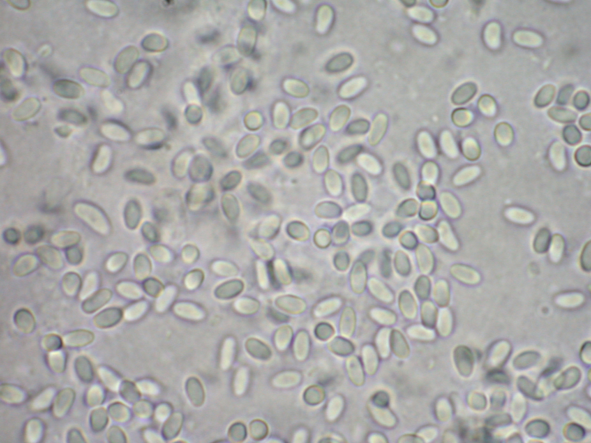
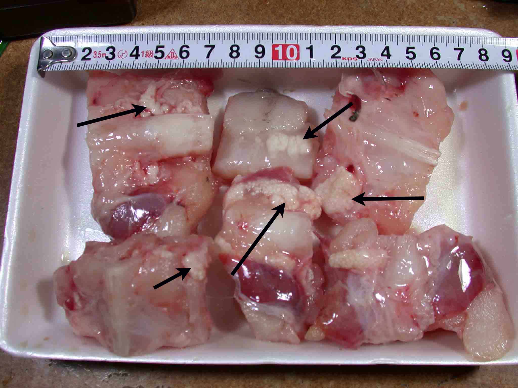
Fig. 2. Xenomas (arrows) found in pieces of anglerfish meat which were sold as 'a set of anglerfish pot' at a supermarket.