| Parasite | Thelohanellus kitauei |
|---|---|
| Taxonomy | Myxozoa, Myxosporea, Bivalvulida |
| Host | Common carp (Cyprinus carpio) |
| Disease name | Intestinal thelohanellosis |
| Infection site | Intestine |
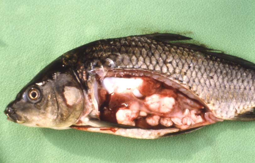
| Clinical signs | This disease occurs only in 1-year fish. Diseased fish exhibit growth retardation, emaciation and abdominal swelling. Many cysts (several mm to several cm in diameter) are observed in the intestine (Fig. 1). |
| Parasitology | Numerous spores are produced inside the tubular vegetative form (Egusa and Nakajima, 1981). A pyriform spore (length 23-29 (average 26.3) mm; width 8-11 (9.2) mm) has one polar capsule (length 14-18 (16.8) mm). A spore is surrounded by a membranous envelope, and the total length is 31-35 (33.4) mm (Fig. 2). The life cycle is unknown. The alternate host is probably involved in the life cycle. |
| Pathology | Tumor-like cysts in the intestinal wall block the intestinal lumen, leading to starvation of the host. Extensive pathological changes are not observed in the peripheral intestinal tissues, but the neighboring hepatopancreas is atrophied due to compression of the expanded intestine. |
| Health hazard | Since this parasite doesn't infect to human, there is no problem in food hygiene |
| Diagnosis | Check the spores by wet-mount of cysts. Sample should be smeared and stained by Giemsa or Diff-Quik. |
| Other information | This disease was first reported from carp farms in Kagoshima Prefecture, the south-western part of Japan, in 1978 and then spread to other districts such as Ibaraki and Nagano Prefecture, the central part of Japan. According to Egusa and Nakajima (1981), the prevalence and the mortality were 22.6 % and 9 %, respectively. However, the disease outbreak decreased after then, and there are no reports in these days. |
| References | Egusa, S. and K. Nakajima (1981): A new myxozoa Thelohanellus kitauei, the cause of intestinal giant cystic disease of carp. Fish Pathol., 15, 213-218. |
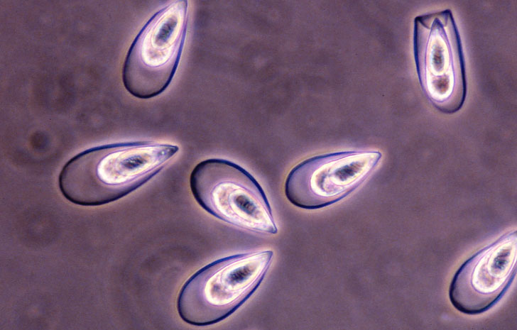

Fig. 1. Common carp showing the intestinal thelohanellosis.
(Photos by S. Egusa (1) and K. Ogawa (2))
Fig. 2. Fresh spores of T. kitauei