| Parasite | Wedlia orientalis |
|---|---|
| Taxonomy | Plathyhelminthes, Trematoda, Didymozoidae |
| Host | Northern bluefin tuna (Thunnus thynnus) |
| Infection site | Gastric submucosa |
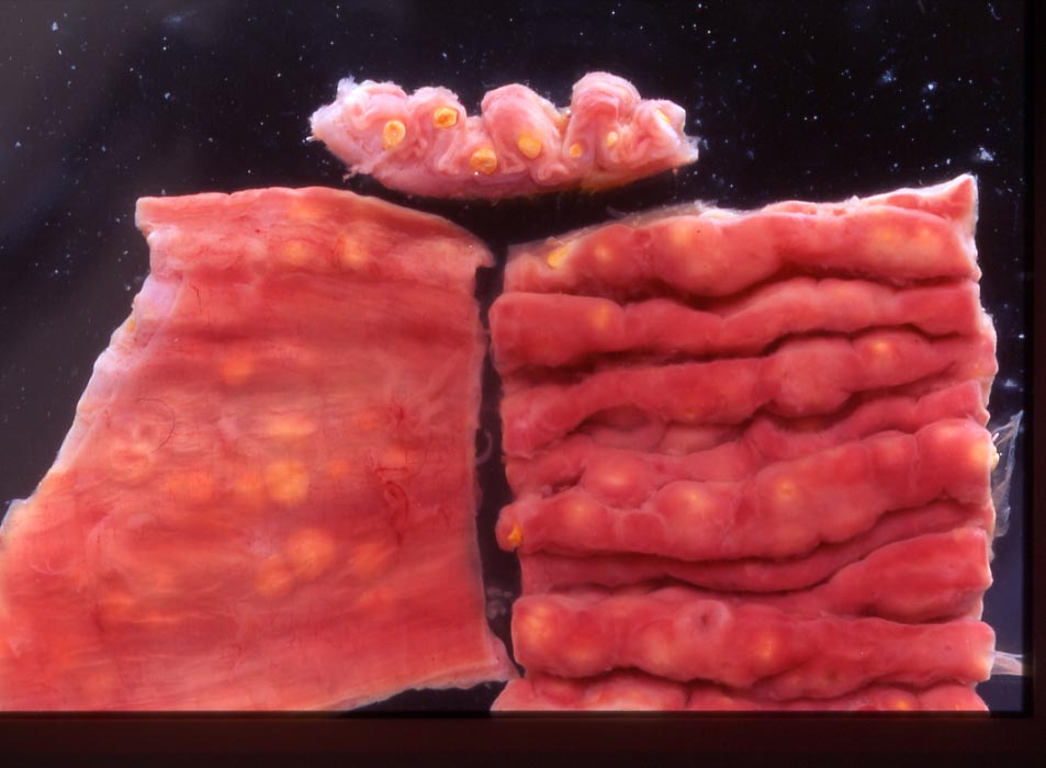
| Clinical sign | Yellowish cysts are observed inside the stomach (Fig. 1). |
| Parasitology | A cyst includes 2 adults, one is male (smaller one) and the other is female. Both worms have an elongated shallow scoop-like forebody with flat apical end (Fig. 2). The hindbody of female is globular. It is partly concaved and lodges the hind body of male (Fig. 2). |
| Pathology | Intensity of infection is not correlated with the growth of fish or BMI, suggesting that the pathogenicity of this parasite is low (Momoyama and Kobayashi, 2004). |
| Health hazard | Since this parasite is not infectious to human, it is harmless in food hygiene. |
| Diagnosis | Check the cysts in the gastric submucosa. |
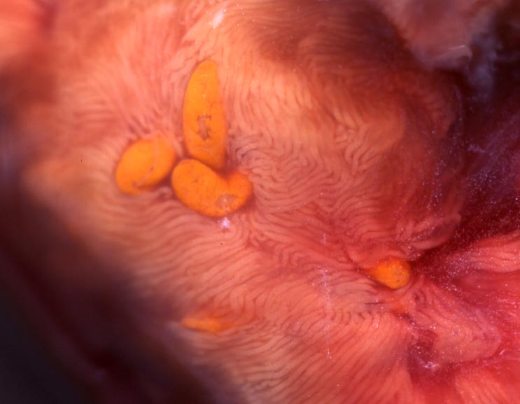
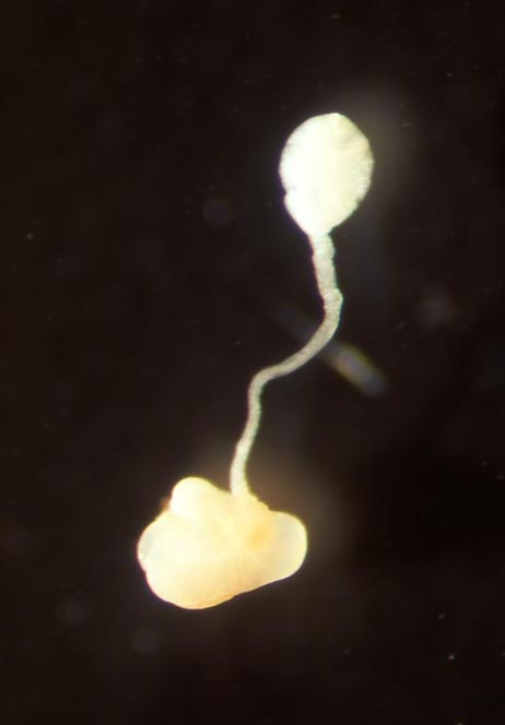
| Other information | Some other didymozooidae trematodes were reported in Northern bluefin tuna, Coeliotrema thynnus in the pyloric caecum (Figs. 3, 4); Dydimocystis wedli in the gill filament; Didymocystis semiglobularis in the epidermis of the gill arch; Didymocystis sloeiformis in the bone tissue of the gill arch; Kollikeria reniformis at the surface of the gill raker. |
| References | Momoyama, K. and T. Kobayashi (2004): Didymozoid
parasites found in the blue fin tuna,
Thunnus thynnus, caught in the coastal waters along Yamaguchi Prefecture in the Japan
Sea.
Bull. Yamaguchi Pref. Fish. Res. Ctr., 2, 125-132. |
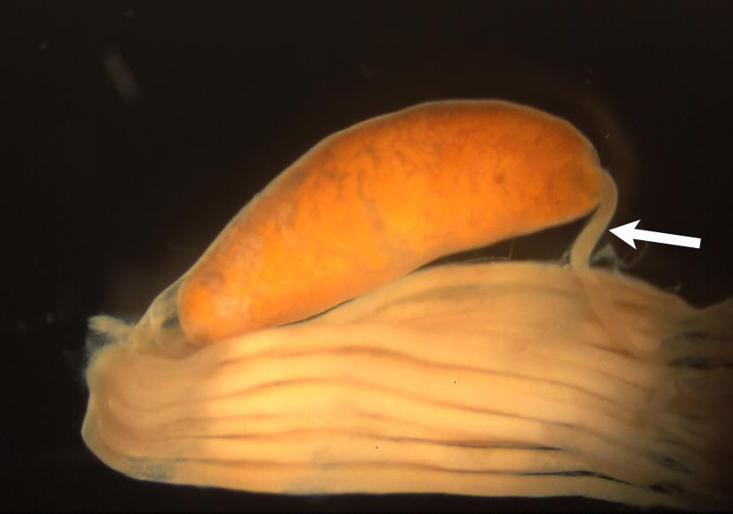
Fig. 4. One string of paluric caecum (arrow), penetrating
through the cyst of C. thynnus.
Fig. 3. Cysts ofC. thynnus in the pyloric caecum.
(Photos by K. Momoyama)
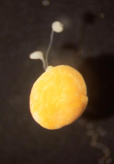
Fig. 1. Cyst of W. orientalis (yellow ones) in the stomach wall.

Fig. 2. Female (left) and male (right) of W. orientalis