(Photos by Mark Freeman (2-4))
| Parasite | X-cell |
|---|---|
| Taxonomy | Protozoa |
| Hosts | Yellowfin goby (Acanthogobius flavimanus), cresthead flounder (Pleuronectes schrenki), flathead flounder (Hippoglossoides dubius), Pacific cod (Gadus macrocephalus), Atlantic cod (Gadus morhua) |
| Infection site | Skin and eye in yellowfin goby, pseudobranch in cods, gill and skin in flatfishes |
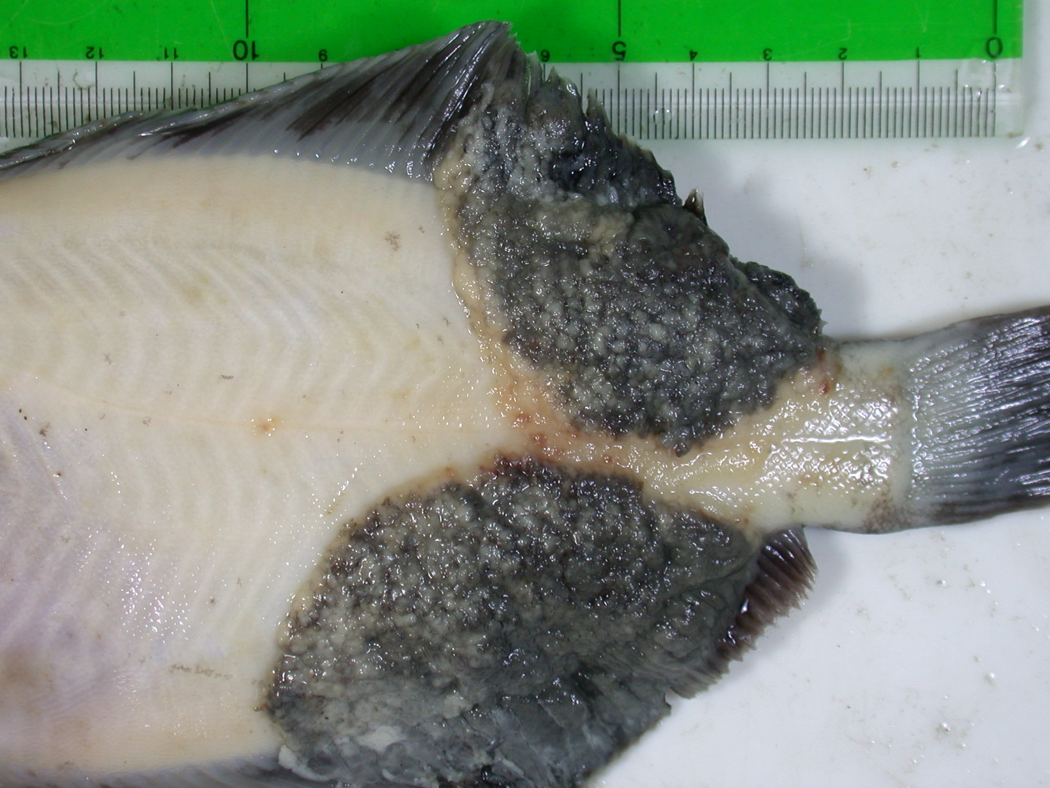
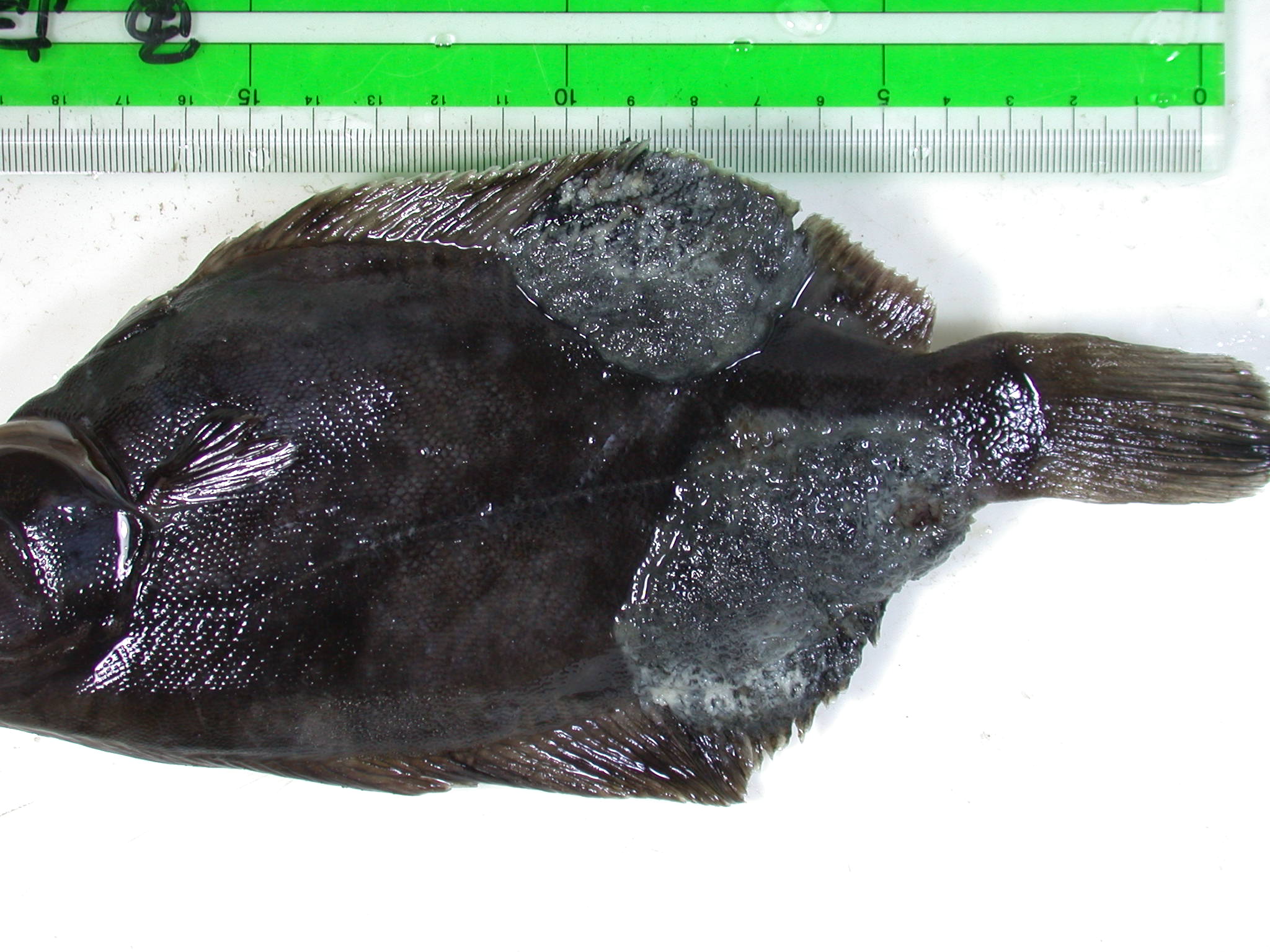
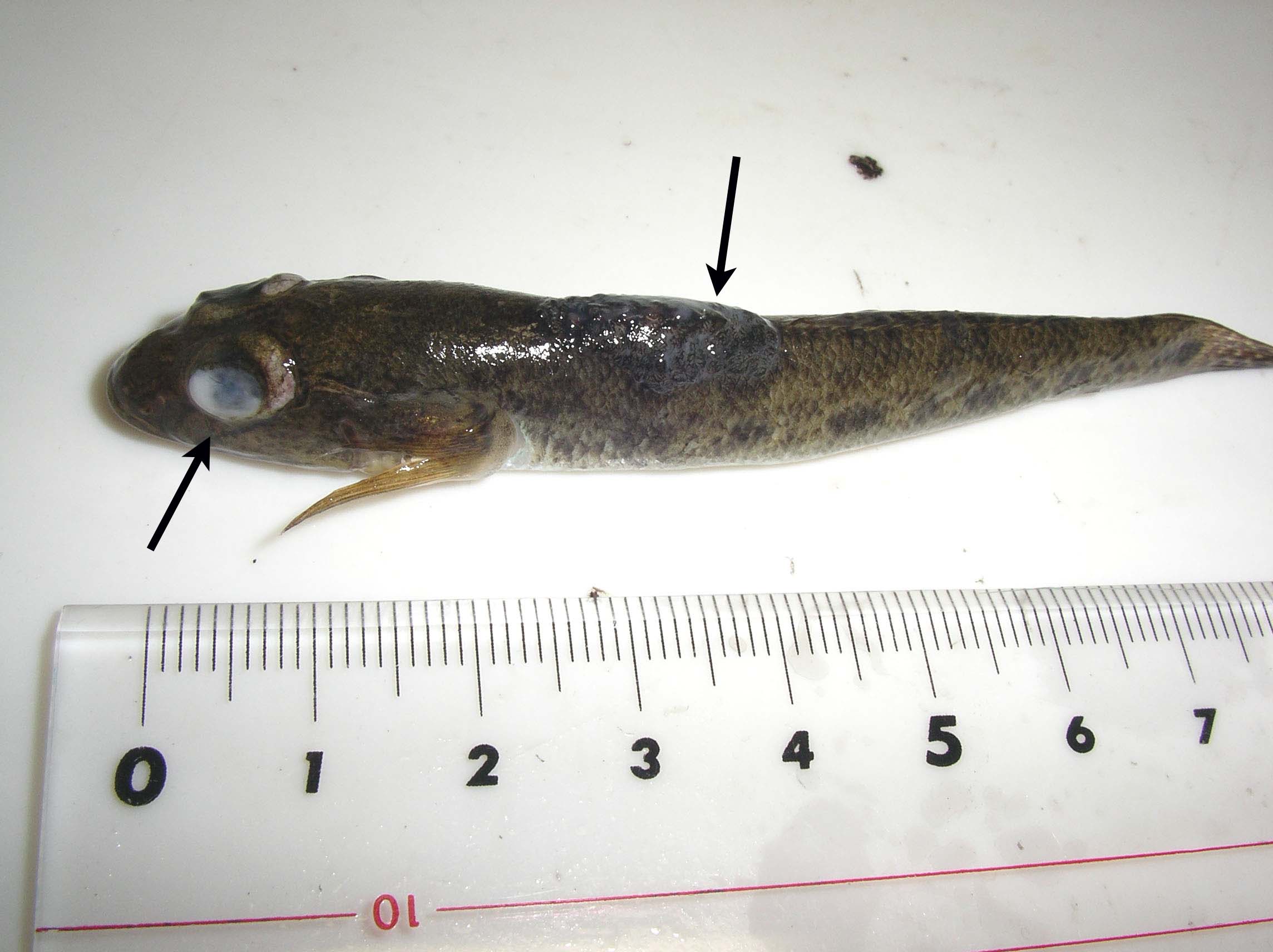
| Clincal sign | Infected yellowfin goby exhibits bulges in the head and exophthalmus (Fig. 1). Infected goby is called as ‘ghost goby’ due to its ugly-looking. Tumor-like lesions are observed (Fig. 2). |
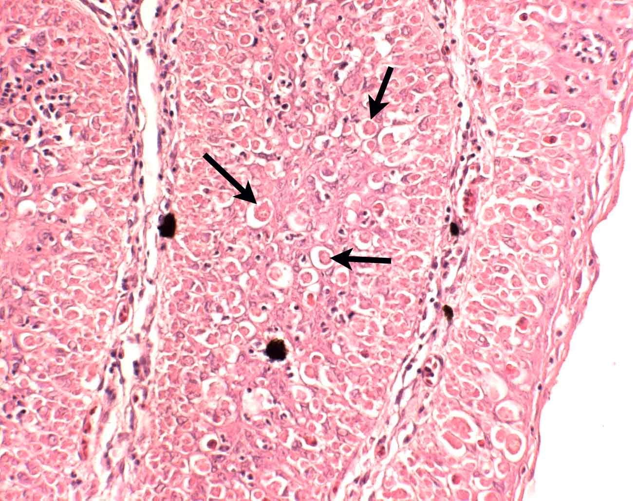
| Parasitology | The causative agent of this disease is called as ‘X-cell’. X-cell (5-20 mm) has a large round nucleus. Molecular analysis indicated that X-cell is a parasitic protozoan though the systematic position has not been clarified yet (Miwa et al., 2004). |
| Pathology | X-cell has a consistent morphology regardless of host species. Tumor-like lesions are formed by its proliferation in the host’s tissue. It is unknown whether this parasite causes a mortality of infected fish. |
| Health hazard | Since this parasite is not infectious to human, it is harmless in food hygiene. |
| Diagnosis | X-cells can be observed under the histological section of the lesions. |
| Other information | This disease is often observed in the bottom-dwelling teleosts, particularly flatfishes, cod or goby living in temperate to cold seawater. Previously, the tumor-like lesions of the disease had been suspected to be caused by water pollution or virus (Shinkawa and Yamazaki, 1987). However, X-cell has now been demonstrated to be a parasitic protozoan. |
| References | Miwa, S., C. Nakayasu,
T. Kamaishi and Y. Yoshiura (2004): X-cells in fish pseudotumors are parasitic
protozoans. Dis. Aquat. Org., 58, 165-170. Shinkawa, T. and F. Yamazaki (1987): Proliferative patterns of X-cells found in the tumorous lesions of Japanese goby. Nippon Suisan Gakkaishi, 53, 563-568. |
Fig. 4. Histological section of X-cells (arrows) in the lesion of
cresthead flounder.
Fig. 3. Tumor-like lesions of cresthead flounder.
Fig. 2. A cresthead flounder showing X-cell disease.
Fig. 1. Lesions (arrows) caused by X-cells in yellowfin goby.
