Fig. 2. The muscle with degenerated lipids.
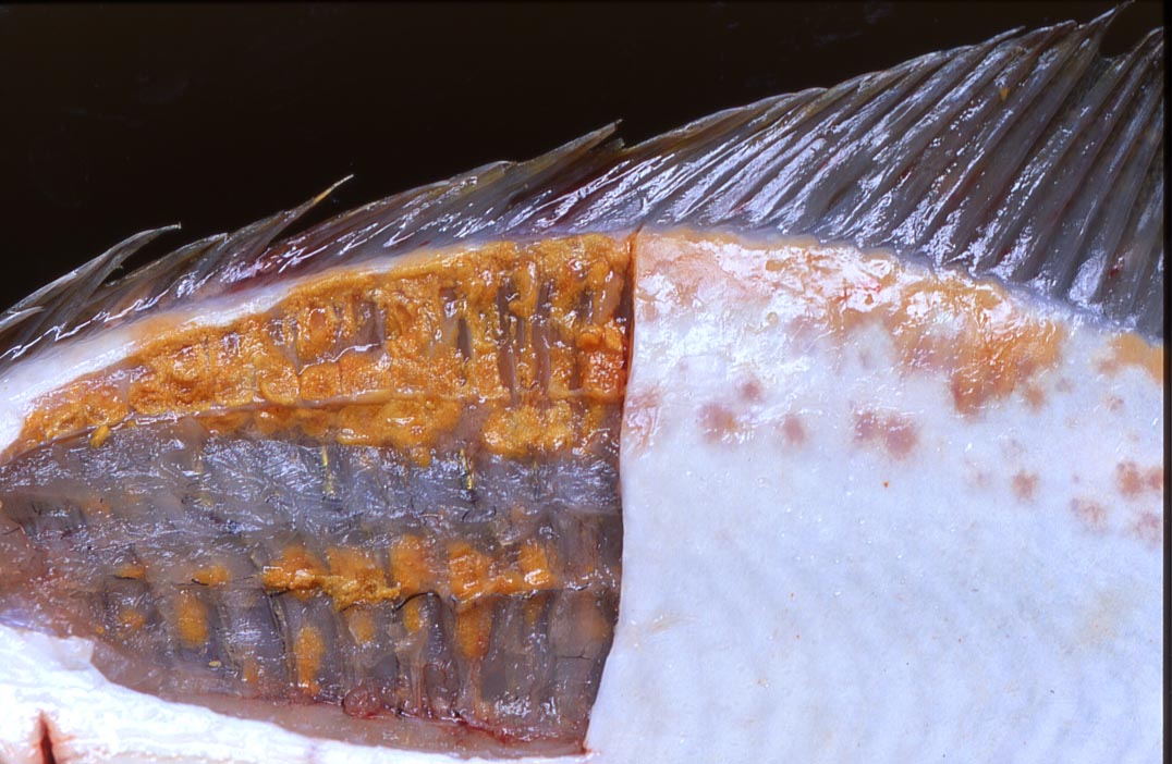

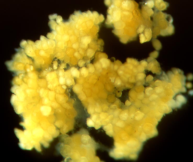
Fig. 3. Degenerated adipose cells.
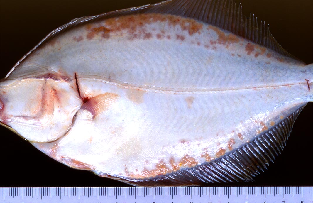
Fig. 1. Blind side of the stone flounder showing many orange patches.
(Photos by K. Momoyama)
| Disease name | Fatty degeneration |
|---|---|
| Fish | Stone flounder (Kareius bicoloratus) |
| Clinical sign | Orange patches are externally observed in the fin muscle of the blind side (Fig. 1). The muscle changes into yellow due to the excessive accumulations of the degenerated lipids (Fig. 2). |
| Pathology | Many yellow to orange adipose cells (70-300 mm) are observed as granules (Fig. 3). Degenerated adipose cells are oval to ellipsoid and harder than normal adipose cells. The cells have a cratered surface, and often include spherical and spiny spherical bodies. Degenerated lipids are stained blue by Sudan Black B and sparingly soluble or insoluble. The cause of the fatty degeneration is unknown. |
| Health hazard | Influences to human by consumption of degenerated lipid are not known. |
| Diagnosis | Degenerated adipose cells are stained by Sudan Black B. |
| Other information | It was reported that diseased fish are found only in winter (Momoyama and Tensha, 2006). Yellow fat disease in cultured fish is caused by the degenerated foods. |
| References | Momoyama, K. and K. Tensha (2006): Ugly-looking parasitic infections and
other abnormalities of wild fish and shellfish caught in the coastal or
inland waters around or in Yamaguchi Prefecture. Bull. Yamaguchi Pref. Fish. Res. Ctr.,
4, 143-161. (In Japanese) |