| Pathogen | Unidentified fungus |
|---|---|
| Taxonomy | Fungi |
| Host | Japanese mantis shrimp (Oratosquilla oratoria) |
| Infection site | Gill, pleopod |
| Clinical sign | Lesions become brown to black in colour due to the necrosis (Fig. 1). |
| Mycology | Branching hyphae are 2.5-5.0 μm in diameter (Fig. 2). A spore is cylindrical and ca. 4.2-12.5 μm in size (Fig. 3). |
| Pathology | It is known that infected Japanese mantis shrimp die after the catch. Pathogenicity of the fungus to the host shrimp may be high since host’s gills are observed to be detached by hyphae infection (Momoyama and Tensha, 2006). |
| Health hazard | Since this fungus is not infectious to human, it is harmless in food hygiene. |
| Diagnosis | No methods for definitive diagnosis are available, because this fungus has not been identified at the species level. |
| References | Momoyama, K. and K. Tensha (2006): Ugly-looking parasitic infections and other abnormalities of wild fish and shellfish caught in the coastal or inland waters around or in Yamaguchi Prefecture. Bull. Yamaguchi Pref. Fish. Res. Ctr., 4, 143-161. (In Japanese) |
Fig. 1. Lesions (arrows) caused by the fungus in Japanese mantis shrimp.
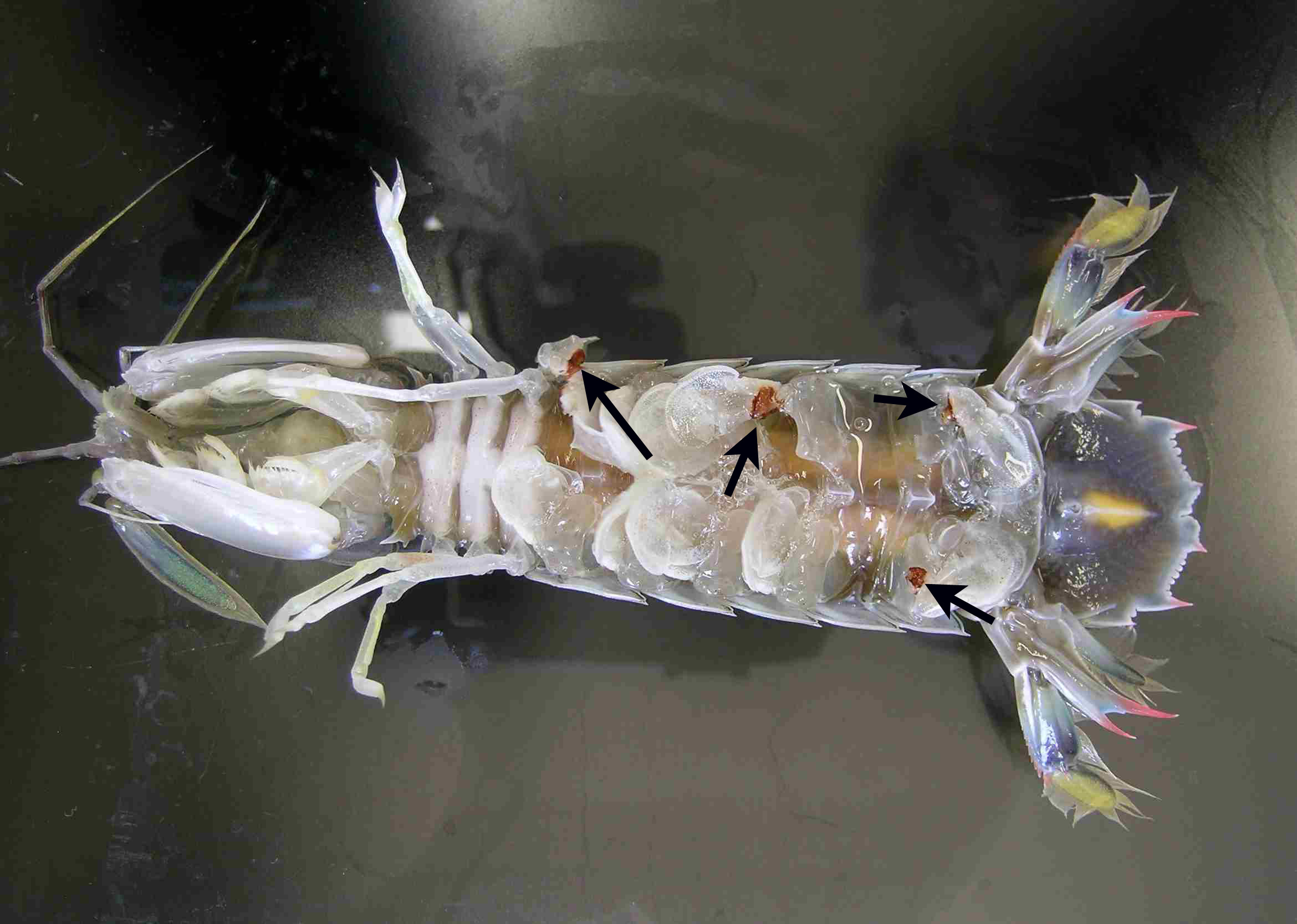
(Photos by K. Tensha and K. Momoyama)
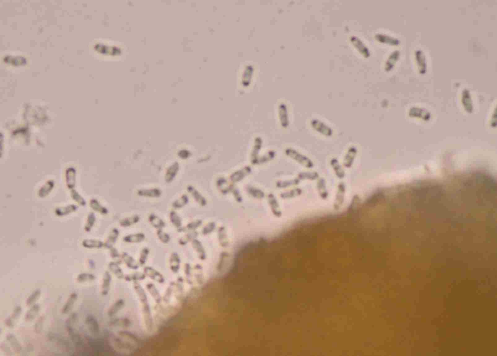
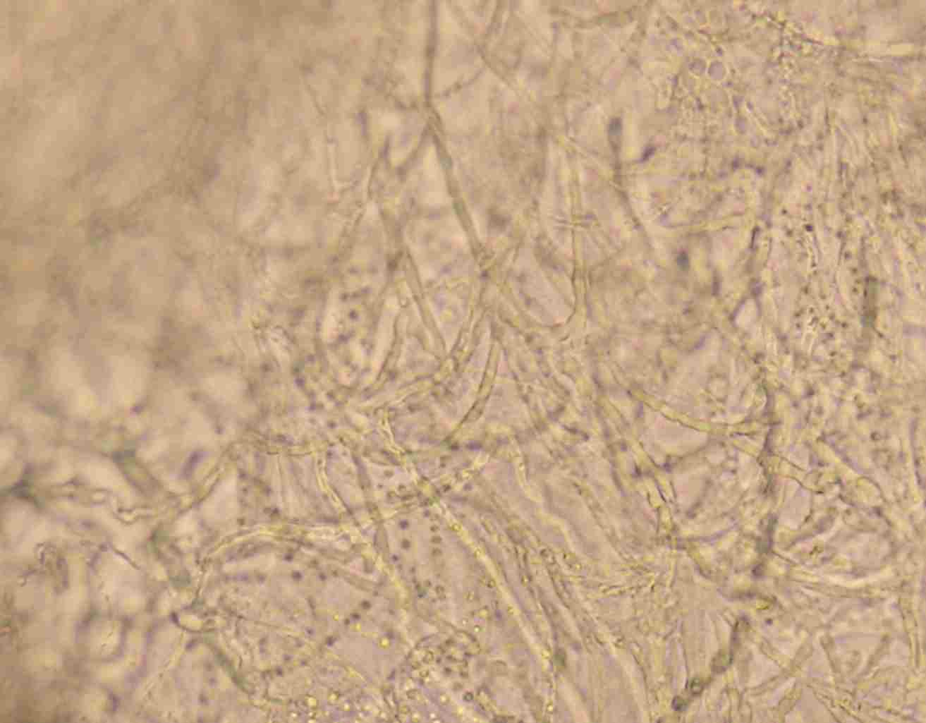
Fig. 3. Spores of the fungus.
Fig. 2. Hyphae in the lesion.