| Parasite | Neobrachiella hugu |
|---|---|
| Taxonomy | Arthropoda, Crustacea, Copepoda, Lernaeopodida |
| Host | Tiger puffer (Takifugu rubripes) |
| Infection site | Buccal cavity wall, rarely around the mouth |
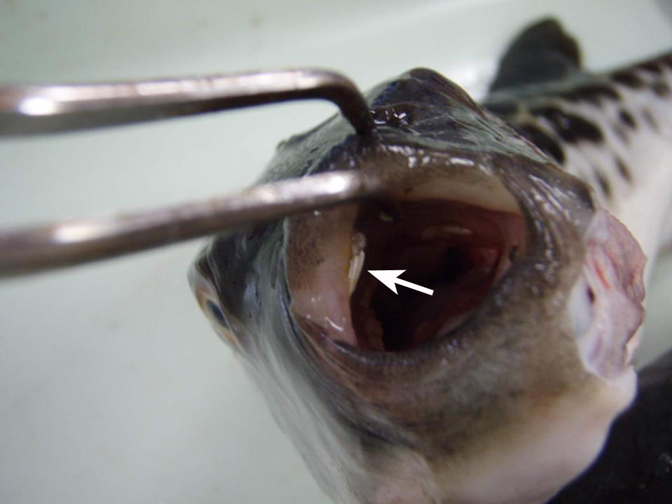
| Clinical sign | White parasites are observed in the buccal cavity (Fig. 1). In case of heavy infection, parasites are seen around the mouth (Fig. 2). |
| Parasitology | Visually observed adults are females, which attach to the host’s tissues by a unique holdfast, known as the ‘bulla’. Cephalothorax is elongate, 3.1-3.6 mm in length. A pair of long egg strings (5.0-6.0 mm in length) is possessed at the posterior end (Fig. 3). This parasite was originally described as Clavellopsis hugu Yamaguti, 1939, and renamed as N. hugu by Ogawa & Inouye (1997). |
| Pathology | Pathogenicities of the parasites are low though the local hyperplasia was provoked at the attachment site. |
| Health hazard | Since this parasite is not infectious to human, it is harmless in food hygiene. |
| Diagnosis | Fix the adult parasite in 70% ethanol and observe in lactic acid. |
| Other information | During the 2-year survey of the parasite in cultured tiger puffer at Nagasaki prefecture, high prevalence of infection (max 100%) was observed throughout the study period but the intensity of infection was as low as 5.6-6.7 parasites per fish. Thus, harmful effects of this parasite were considered to be negligible (Ogawa and Inouye, 1997). |
| References | Ogawa, K. and K.
Inouye (1997): Parasites of cultured tiger puffer (Takifugu rubripes) and their seasonal occurrences, with
descriptions of two new species of Gyrodactylus.
Fish Pathol., 32, 7-14. Yamaguti, S. (1939): Parasitic copepods from fishes of Japan. Part 6. Lernaeopodida, I. Vol. Jub. Prof. Yoshida, 2, 529-578. |
Fig. 1. Neobrachiella hugu (arrow) parasitizing in the buccal cavity of tiger puffer.
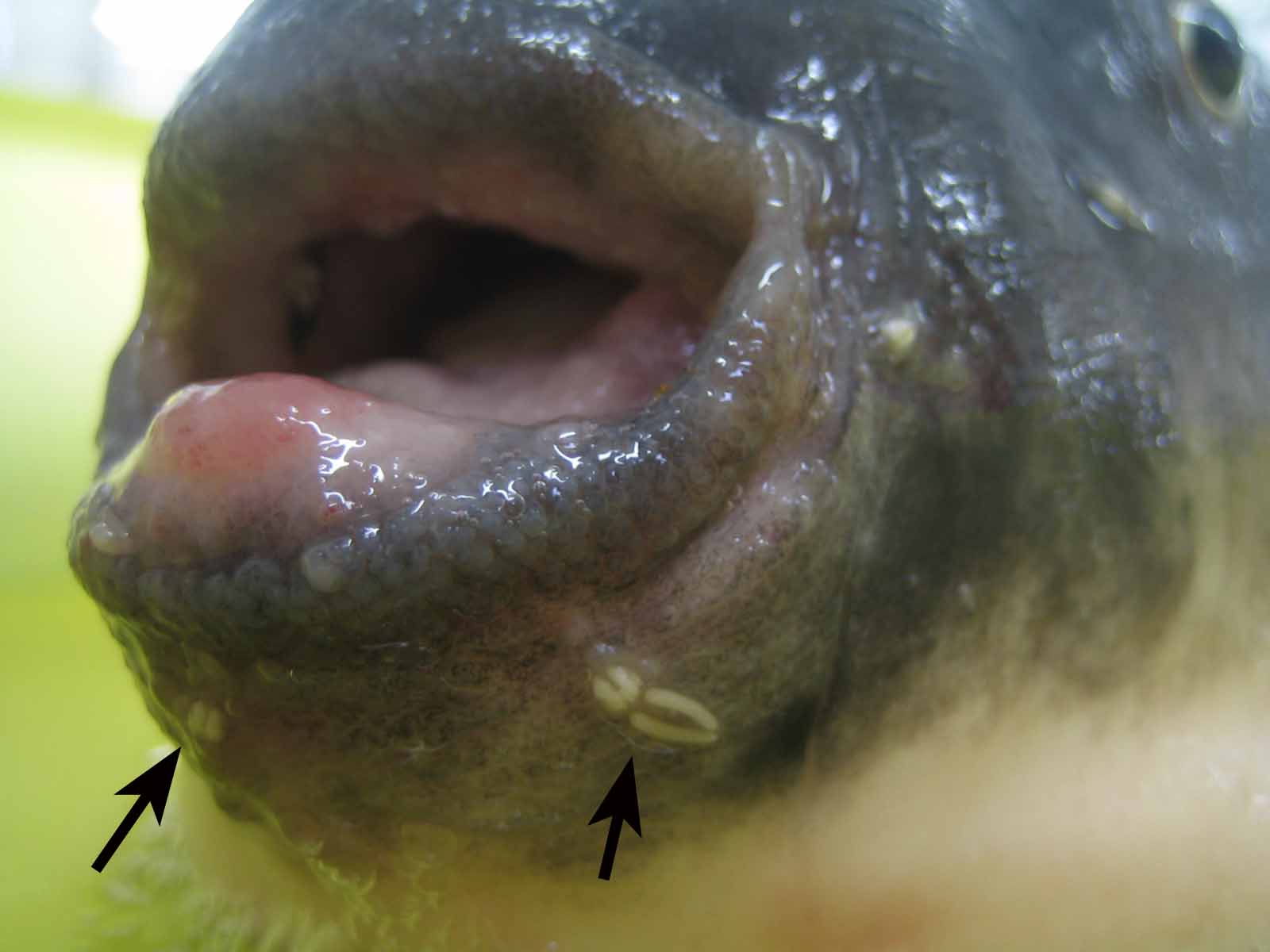
(Photos by H. Donai (1, 3) , R. Kinami (2))

Fig. 3. Neobrachiella hugu collected from the fish.
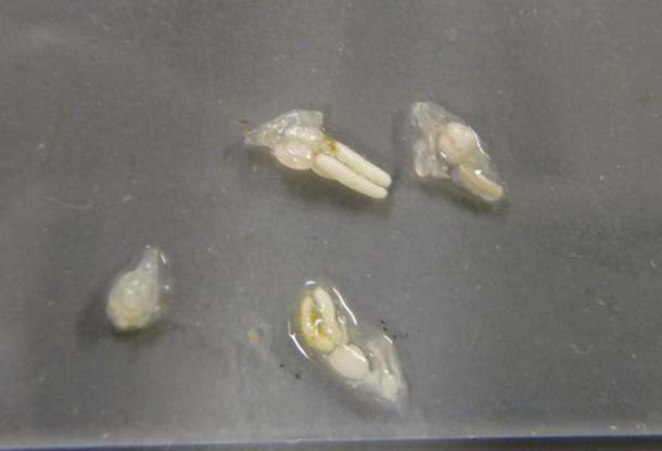
Fig. 2. N. hugu parasitizing around the mouth (arrows).